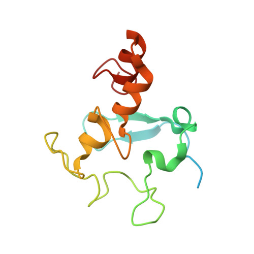
2A3M

Structure of Desulfovibrio desulfuricans G20 tetraheme cytochrome (oxidized form)


Experimental Data Snapshot

  • Method: X-RAY DIFFRACTION
  • Resolution: 1.50 Å
  • R-Value Free: 0.195 
  • R-Value Work: 0.175 
  • R-Value Observed: 0.177 

Starting Model: experimental
View more details

wwPDB Validation   3D Report Full Report


Ligand Structure Quality Assessment 

Created with Raphaël 2.3.0Worse 01 BetterLigand structure goodness of fit to experimental dataBest fitted HECClick on this verticalbar to view details

This is version 2.2 of the entry. See complete history


Literature

Desulfovibrio desulfuricans G20 Tetraheme Cytochrome Structure at 1.5A and Cytochrome Interaction with Metal Complexes

Pattarkine, M.V.Tanner, J.J.Bottoms, C.A.Lee, Y.H.Wall, J.D.

(2006) J Mol Biol 358: 1314-1327

  • DOI: https://doi.org/10.1016/j.jmb.2006.03.010
  • Primary Citation of Related Structures:  
    2A3M, 2A3P

  • PubMed Abstract: 

    The structure of the type I tetraheme cytochrome c(3) from Desulfovibrio desulfuricans G20 was determined to 1.5 Angstrom by X-ray crystallography. In addition to the oxidized form, the structure of the molybdate-bound form of the protein was determined from oxidized crystals soaked in sodium molybdate. Only small structural shifts were obtained with metal binding, consistent with the remarkable structural stability of this protein. In vitro experiments with pure cytochrome showed that molybdate could oxidize the reduced cytochrome, although not as rapidly as U(VI) present as uranyl acetate. Alterations in the overall conformation and thermostability of the metal-oxidized protein were investigated by circular dichroism studies. Again, only small changes in protein structure were documented. The location of the molybdate ion near heme IV in the crystal structure suggested heme IV as the site of electron exit from the reduced cytochrome and implicated Lys14 and Lys56 in binding. Analysis of structurally conserved water molecules in type I cytochrome c(3) crystal structures identified interactions predicted to be important for protein stability and possibly for intramolecular electron transfer among heme molecules.


  • Organizational Affiliation

    Biochemistry Department, University of Missouri-Columbia, Columbia, MO 65211, USA.


Macromolecules
Find similar proteins by:  (by identity cutoff)  |  3D Structure
Entity ID: 1
MoleculeChains Sequence LengthOrganismDetailsImage
COG3005: Nitrate/TMAO reductases, membrane-bound tetraheme cytochrome c subunit130Oleidesulfovibrio alaskensis G20Mutation(s): 0 
UniProt
Find proteins for Q30WH0 (Oleidesulfovibrio alaskensis (strain ATCC BAA-1058 / DSM 17464 / G20))
Explore Q30WH0 
Go to UniProtKB:  Q30WH0
Entity Groups  
Sequence Clusters30% Identity50% Identity70% Identity90% Identity95% Identity100% Identity
UniProt GroupQ30WH0
Sequence Annotations
Expand
  • Reference Sequence
Experimental Data & Validation

Experimental Data

  • Method: X-RAY DIFFRACTION
  • Resolution: 1.50 Å
  • R-Value Free: 0.195 
  • R-Value Work: 0.175 
  • R-Value Observed: 0.177 
  • Space Group: P 21 21 21
Unit Cell:
Length ( Å )Angle ( ˚ )
a = 43.67α = 90
b = 43.876β = 90
c = 57.729γ = 90
Software Package:
Software NamePurpose
DENZOdata reduction
SCALEPACKdata scaling
REFMACrefinement
PDB_EXTRACTdata extraction

Structure Validation

View Full Validation Report


Ligand Structure Quality Assessment 

Created with Raphaël 2.3.0Worse 01 BetterLigand structure goodness of fit to experimental dataBest fitted HECClick on this verticalbar to view details

Entry History 

Deposition Data

Revision History  (Full details and data files)

  • Version 1.0: 2006-04-25
    Type: Initial release
  • Version 1.1: 2008-04-30
    Changes: Version format compliance
  • Version 1.2: 2011-07-13
    Changes: Advisory, Source and taxonomy, Version format compliance
  • Version 1.3: 2017-10-11
    Changes: Refinement description
  • Version 2.0: 2021-03-03
    Changes: Atomic model, Data collection, Derived calculations, Non-polymer description, Structure summary
  • Version 2.1: 2023-08-23
    Changes: Data collection, Database references, Refinement description
  • Version 2.2: 2024-11-06
    Changes: Structure summary