29YD | pdb_000029yd

Cryo-EM structure of Rhodobacter capsulatus cytochrome bc1 dimer with one Rieske protein in the c position and one in an intermediate position


Experimental Data Snapshot

  • Method: ELECTRON MICROSCOPY
  • Resolution: 2.73 Å
  • Aggregation State: PARTICLE 
  • Reconstruction Method: SINGLE PARTICLE 

Starting Model: experimental
View more details

wwPDB Validation 3D Report Full Report

Validation slider image for 29YD

This is version 1.0 of the entry. See complete history

Literature

Distinct ubiquinone binding at the oxidation and reduction sites of cytochrome bc 1.

Pietras, R.Wojcik-Augustyn, A.Mielecki, B.Sarewicz, M.Jaciuk, M.Koziej, L.Glatt, S.Osyczka, A.

(2026) Proc Natl Acad Sci U S A 123: e2618242123-e2618242123

  • DOI: https://doi.org/10.1073/pnas.2618242123
  • Primary Citation Related Structures: 
    29XZ, 29YA, 29YB, 29YC, 29YD, 29YE

  • PubMed Abstract: 

    The function of cytochrome bc 1 , a widespread energy-conserving enzyme, requires the coordinated activity of two quinone-binding sites (Q o catalyzing oxidation of ubiquinol and Q i catalyzing reduction of ubiquinone). The operation of Q o , but not Q i , involves large-scale movement of the head domain of iron-sulfur protein (ISP-HD). How the respective sites accommodate quinone molecules for efficient catalysis remains elusive. Here, we present high-resolution cryoelectron microscopy structures of bacterial cytochrome bc 1 with native ubiquinone molecules in various states. They show that the quinone headgroup occupies a catalytically competent position in Q o only when the ISP-HD interacts with cytochrome b. When the ISP-HD does not interact with this subunit, quinone is present in the hydrophobic groove, however its headgroup is prevented from reaching the catalytic cavity by steric hindrance. In this state, the position of quinone headgroup is clearly not fixed. In contrast, all structures show Q i in the same state with a well-resolved and catalytically competent quinone headgroup, but with its tail not fixed. These distinctly different ubiquinone binding modes for Q o and Q i secure the smooth operation of cytochrome bc 1 .


  • Organizational Affiliation
    • Faculty of Biochemistry, Biophysics and Biotechnology, Department of Molecular Biophysics, Jagiellonian University, Kraków 30-387, Poland.

Macromolecule Content 

  • Total Structure Weight: 206.44 kDa 
  • Atom Count: 13,978 
  • Modeled Residue Count: 1,708 
  • Deposited Residue Count: 1,784 
  • Unique protein chains: 3

Macromolecules

Find similar proteins by:|  3D Structure
Entity ID: 1
MoleculeChains  Sequence LengthOrganismDetailsImage
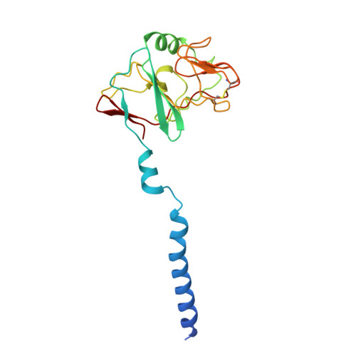
Ubiquinol-cytochrome c reductase iron-sulfur subunit
A, D
190Rhodobacter capsulatus SB 1003Mutation(s): 0 
Gene Names: petAfbcFRCAP_rcc02768
EC: 7.1.1.8
UniProt
Find proteins for D5ANZ2 (Rhodobacter capsulatus (strain ATCC BAA-309 / NBRC 16581 / SB1003))
Explore D5ANZ2 
Go to UniProtKB:  D5ANZ2
Entity Groups
Sequence Clusters30% Identity50% Identity70% Identity90% Identity95% Identity100% Identity
UniProt GroupD5ANZ2
Sequence Annotations
Expand
Reference Sequence
Find similar proteins by:|  3D Structure
Entity ID: 2
MoleculeChains  Sequence LengthOrganismDetailsImage
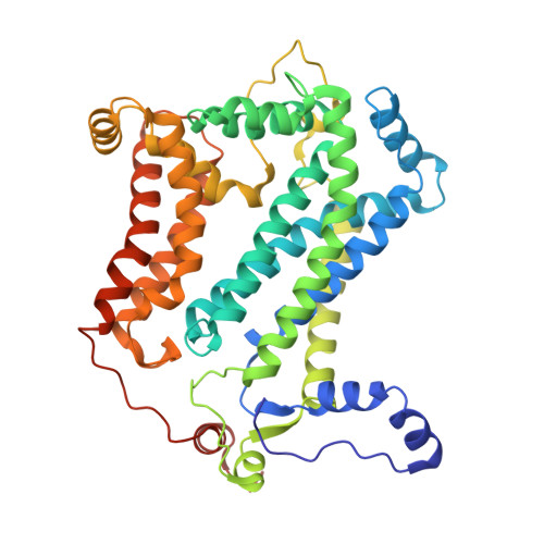
Cytochrome b
B, E
444Rhodobacter capsulatus SB 1003Mutation(s): 0 
Gene Names: petBcytBRCAP_rcc02769
UniProt
Find proteins for D5ANZ3 (Rhodobacter capsulatus (strain ATCC BAA-309 / NBRC 16581 / SB1003))
Explore D5ANZ3 
Go to UniProtKB:  D5ANZ3
Entity Groups
Sequence Clusters30% Identity50% Identity70% Identity90% Identity95% Identity100% Identity
UniProt GroupD5ANZ3
Sequence Annotations
Expand
Reference Sequence
Find similar proteins by:|  3D Structure
Entity ID: 3
MoleculeChains  Sequence LengthOrganismDetailsImage
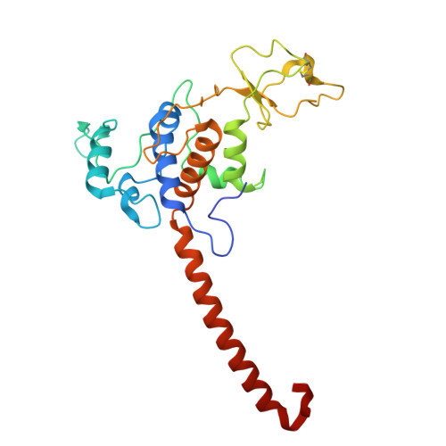
Cytochrome c1
C, F
258Rhodobacter capsulatus SB 1003Mutation(s): 0 
Gene Names: petCRCAP_rcc02770
UniProt
Find proteins for D5ANZ4 (Rhodobacter capsulatus (strain ATCC BAA-309 / NBRC 16581 / SB1003))
Explore D5ANZ4 
Go to UniProtKB:  D5ANZ4
Entity Groups
Sequence Clusters30% Identity50% Identity70% Identity90% Identity95% Identity100% Identity
UniProt GroupD5ANZ4
Sequence Annotations
Expand
Reference Sequence

Small Molecules

Ligands 5 Unique
IDChains Name / Formula / InChI Key2D Diagram3D Interactions
HEC
(Subject of Investigation/LOI)

Query on HEC


Download:Ideal Coordinates CCD File
O [auth C],
X [auth F]
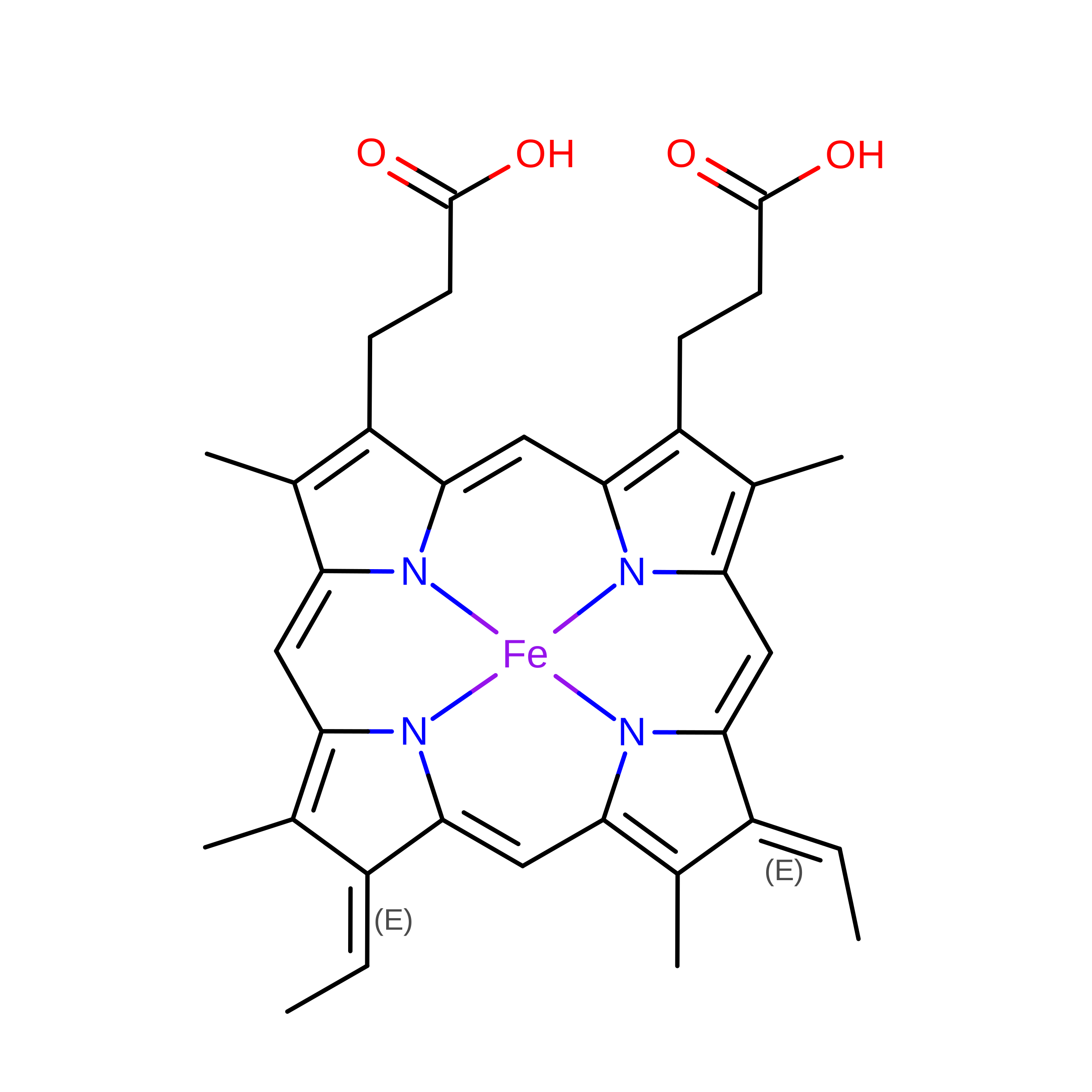
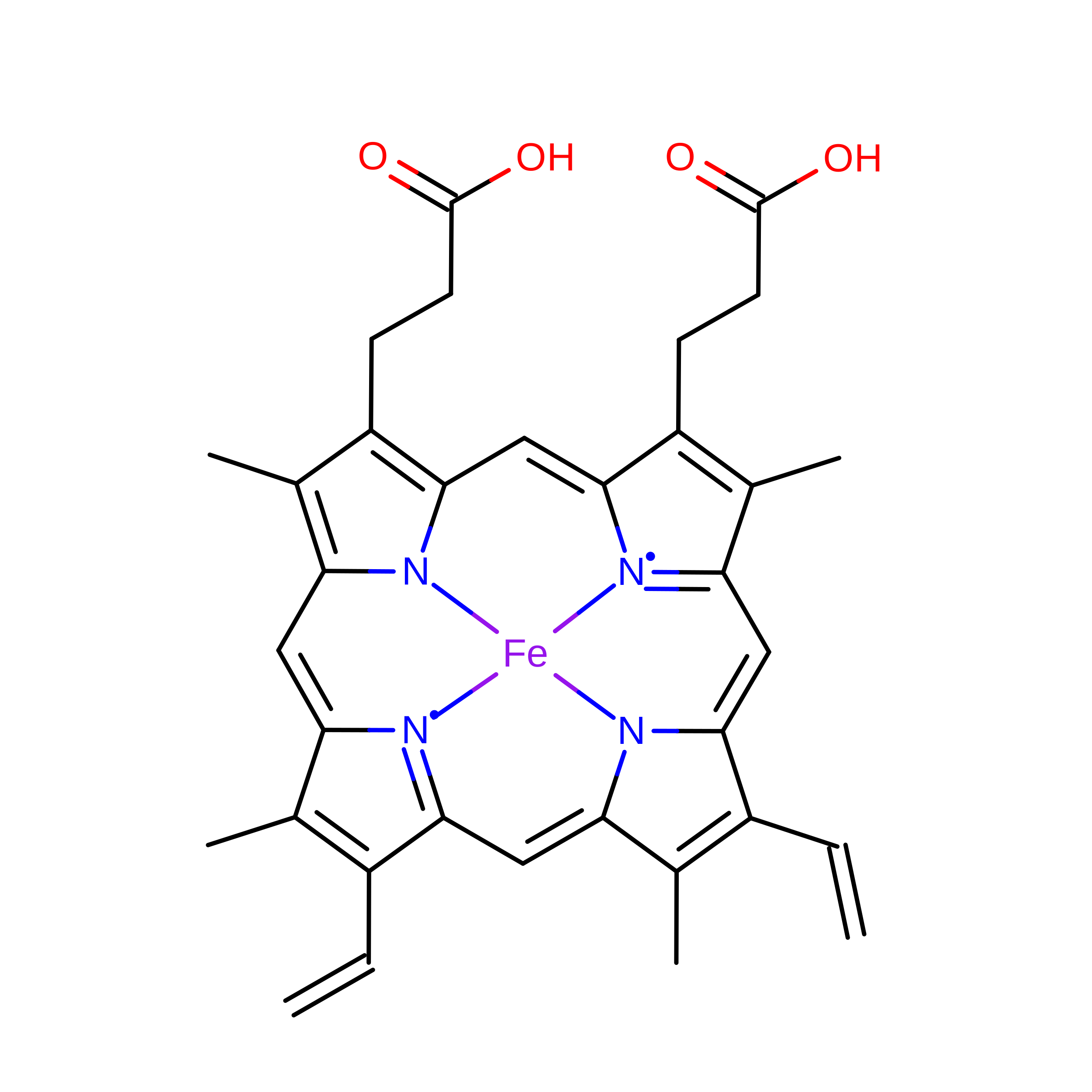
HEME C
C34 H36 Fe N4 O4
YZTICFPLOXKSQO-RGGAHWMASA-L
HEM
(Subject of Investigation/LOI)

Query on HEM


Download:Ideal Coordinates CCD File
I [auth B],
J [auth B],
R [auth E],
S [auth E]
PROTOPORPHYRIN IX CONTAINING FE
C34 H32 Fe N4 O4
KABFMIBPWCXCRK-RGGAHWMASA-L
UMQ

Query on UMQ


Download:Ideal Coordinates CCD File
H [auth A]
L [auth B]
M [auth B]
N [auth B]
Q [auth D]
H [auth A],
L [auth B],
M [auth B],
N [auth B],
Q [auth D],
U [auth E],
V [auth E],
W [auth E]
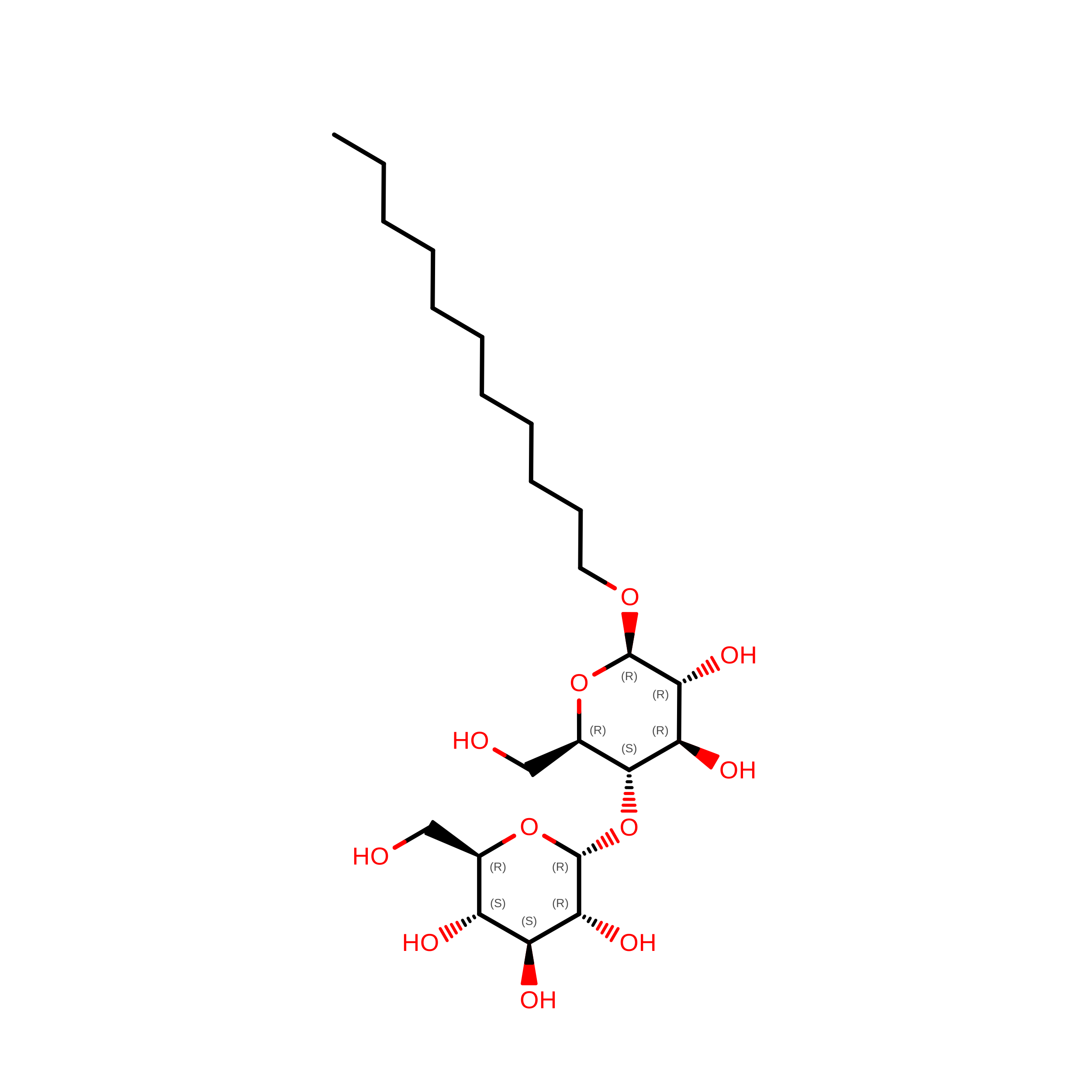
UNDECYL-MALTOSIDE
C23 H44 O11
UYEMNFYVTFDKRG-ZNGNCRBCSA-N
UQ1
(Subject of Investigation/LOI)

Query on UQ1


Download:Ideal Coordinates CCD File
K [auth B],
T [auth E]
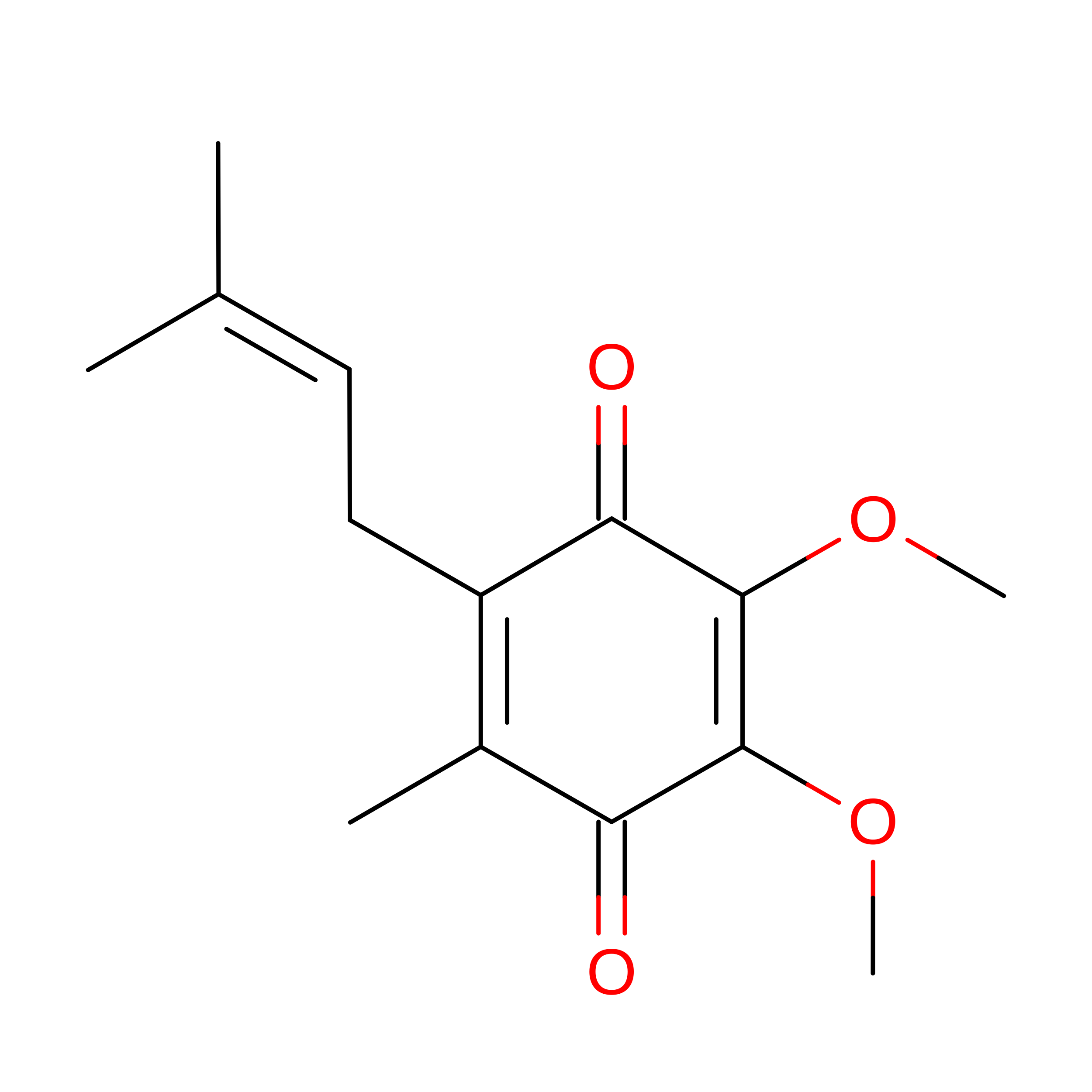
UBIQUINONE-1
C14 H18 O4
SOECUQMRSRVZQQ-UHFFFAOYSA-N
FES
(Subject of Investigation/LOI)

Query on FES


Download:Ideal Coordinates CCD File
G [auth A],
P [auth D]
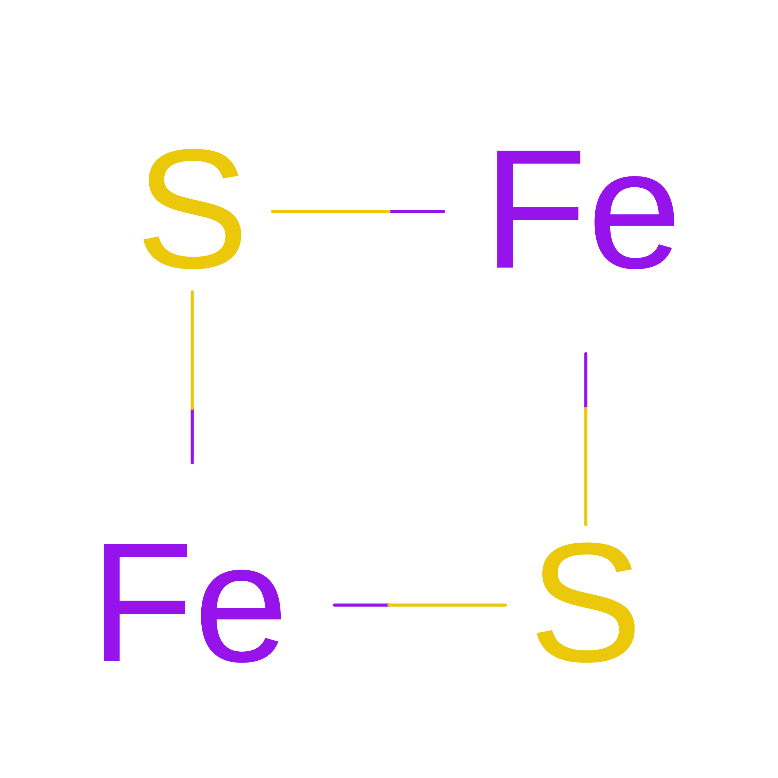
FE2/S2 (INORGANIC) CLUSTER
Fe2 S2
NIXDOXVAJZFRNF-UHFFFAOYSA-N

Experimental Data & Validation

Experimental Data

  • Method: ELECTRON MICROSCOPY
  • Resolution: 2.73 Å
  • Aggregation State: PARTICLE 
  • Reconstruction Method: SINGLE PARTICLE 
EM Software:
TaskSoftware PackageVersion
RECONSTRUCTIONcryoSPARC4.7
MODEL REFINEMENTCoot0.9.8
MODEL REFINEMENTPHENIX2.0

Structure Validation

View Full Validation Report


Entry History 

& Funding Information

Deposition Data


Funding OrganizationLocationGrant Number
Polish National Science CentrePoland2023/49/B/NZ1/02110
Ministry of Science and Higher Education (Poland)Poland1/SOL/2021/2

Revision History  (Full details and data files)

  • Version 1.0: 2026-09-09
    Type: Initial release