Download Mendeley
Discovery and Optimization of Novel Nonhydroxamate LpxC Inhibitors for the Treatment of Multidrug-Resistant Gram-Negative Infections.
Martin, D.P., Teng, M., Nammalwar, B., Perez, C., Li, X., Munguia, J., Taganov, K., Fan, J., Agarwalla, S., Lonergan, D., Tomaras, A.P., Zimmerman, Z., Puerta, D.T.(2026) J Med Chem 69: 18692-18704
- PubMed: 42593942 Search on PubMedSearch on PubMed Central
- DOI: https://doi.org/10.1021/acs.jmedchem.6c01036
- Primary Citation Related Structures:
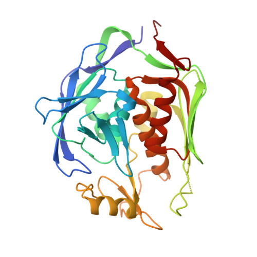
10RY, 29AH, 29AI, 29AK - PubMed Abstract:
This report summarizes the discovery and optimization of a novel series of nonhydroxamate inhibitors targeting LpxC, a Zn2+-dependent hydrolase that is essential for the survival of Gram-negative bacteria. Beginning with a 5-hydroxypyrimidin-4-one metal-binding pharmacophore, structure-based approaches were utilized to generate a series of potent inhibitors that exhibited activity against a wide variety of Enterobacterales, including both susceptible and multidrug-resistant pathogens, and efficacy in murine thigh infection models. These novel compounds were evaluated in a rat model of cardiovascular toxicity to demonstrate the safety of the scaffold relative to another LpxC inhibitor that proved unsuccessful in Phase I clinical trials. A variety of inhibitors with potent in vivo efficacy and no hemodynamic effects were identified, which constituted an initial suite of potential development candidates.
- Blacksmith Medicines, Inc., San Diego, California92121, United States.
Organizational Affiliation: