Download Mendeley
Structural basis of the gating mechanism of the large-conductance mechanosensitive channel from Escherichia coli.
Hardman, K., Wort, J.L., Waheed, Q., Liu, X., Arul, D., Porav, S.A., Calabrese, A.N., Muench, S.P., Pliotas, C.(2026) Nat Commun 17
- PubMed: 42869890 Search on PubMedSearch on PubMed Central
- DOI: https://doi.org/10.1038/s41467-026-76193-0
- Primary Citation Related Structures:
28OA, 28OB, 28OC - PubMed Abstract:
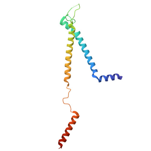
The mechanosensitive channel of large conductance (MscL) is a tension-gated, pore-forming protein that acts as a safety valve to protect bacteria from osmotic lysis. Escherichia coli MscL (EcMscL) was the first mechanosensitive channel discovered and subsequently served as a model system for understanding mechanical sensing, becoming one of the most decorated and well-studied systems. Despite extensive biophysical and functional characterisation spanning several decades, the precise mechanism of EcMscL gating has been poorly understood due to the lack of high-resolution structural information. Herein, we solve two EcMscL structures by cryoEM in the closed conformation in DMPC and DOPC lipid nanodiscs. Using PELDOR/DEER spectroscopy, we screen conditions and identify that in DSPC lipids, the EcMscL conformational ensemble shifts away from the closed state and that open-like states are present. We solve the structure in an expanded state by cryoEM, revealing an architecture with pore properties consistent with previous electrophysiology reports. By combining hydrogen-deuterium exchange mass spectrometry and molecular dynamics simulations, we investigate the dynamics of EcMscL gating in lipid bilayers, identifying sites involved in the closed-to-expanded transition. Combined, this enables us to inform on the elusive structural mechanism of EcMscL mechanosensitive channel function.
- Astbury Centre for Structural Molecular Biology, University of Leeds, Leeds, UK.
Organizational Affiliation: