






 Download Mendeley
Structural basis for the synaptic adhesion protein ELFN1-mediated modulation of mGlu7
Lin, S., Feng, Y., Shui, S., Chen, M., Chu, X., Yi, C., Han, S., Zhao, Q., Wu, B.To be published.
Experimental Data Snapshot
wwPDB Validation
Currently 25TW does not have a validation slider image.
Entity ID: 1 | |||||
|---|---|---|---|---|---|
| Molecule | Chains | Sequence Length | Organism | Details | Image |
| Metabotropic glutamate receptor 7 | 920 | Homo sapiens | Mutation(s): 0 Gene Names: GRM7, GPRC1G, MGLUR7 | 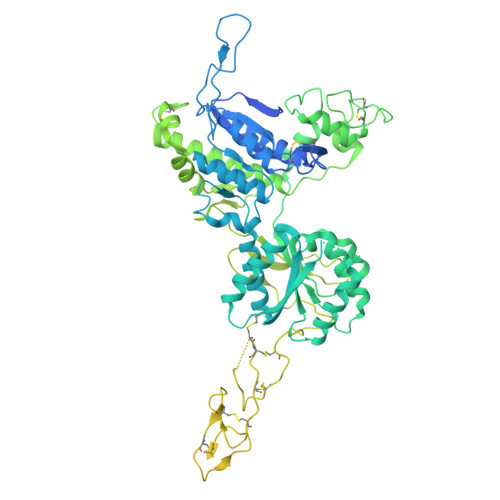 | |
UniProt & NIH Common Fund Data Resources | |||||
PHAROS: Q14831 GTEx: ENSG00000196277 | |||||
Entity Groups | |||||
| Sequence Clusters | 30% Identity50% Identity70% Identity90% Identity95% Identity100% Identity | ||||
| UniProt Group | Q14831 | ||||
Glycosylation | |||||
| Glycosylation Sites: 2 | Go to GlyGen: Q14831-1 | ||||
Sequence AnnotationsExpand | |||||
Reference Sequence | |||||
Entity ID: 2 | |||||
|---|---|---|---|---|---|
| Molecule | Chains | Sequence Length | Organism | Details | Image |
| Protein ELFN1 | 411 | Homo sapiens | Mutation(s): 0 Gene Names: ELFN1, PPP1R28 | 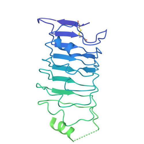 | |
UniProt & NIH Common Fund Data Resources | |||||
PHAROS: P0C7U0 GTEx: ENSG00000225968 | |||||
Entity Groups | |||||
| Sequence Clusters | 30% Identity50% Identity70% Identity90% Identity95% Identity100% Identity | ||||
| UniProt Group | P0C7U0 | ||||
Glycosylation | |||||
| Glycosylation Sites: 3 | Go to GlyGen: P0C7U0-1 | ||||
Sequence AnnotationsExpand | |||||
Reference Sequence | |||||
Entity ID: 3 | |||||
|---|---|---|---|---|---|
| Molecule | Chains | Length | 2D Diagram | Glycosylation | D Interactions |
| beta-D-mannopyranose-(1-3)-2-acetamido-2-deoxy-beta-D-glucopyranose-(1-4)-[alpha-L-fucopyranose-(1-6)]2-acetamido-2-deoxy-beta-D-glucopyranose | E | 4 | N/A | N-Glycosylation | |
Entity ID: 4 | |||||
|---|---|---|---|---|---|
| Molecule | Chains | Length | 2D Diagram | Glycosylation | D Interactions |
| 2-acetamido-2-deoxy-beta-D-glucopyranose-(1-4)-2-acetamido-2-deoxy-beta-D-glucopyranose | F, H, I, J | 2 | N/A | N-Glycosylation | |
Entity ID: 5 | |||||
|---|---|---|---|---|---|
| Molecule | Chains | Length | 2D Diagram | Glycosylation | D Interactions |
| beta-D-mannopyranose-(1-4)-2-acetamido-2-deoxy-beta-D-glucopyranose-(1-4)-2-acetamido-2-deoxy-beta-D-glucopyranose | G | 3 | N/A | N-Glycosylation | |
| Ligands 1 Unique | |||||
|---|---|---|---|---|---|
| ID | Chains | Name / Formula / InChI Key | 2D Diagram | 3D Interactions | |
| NAG Download:Ideal Coordinates CCD File | K [auth A], L [auth A], M [auth B], N [auth B] | 2-acetamido-2-deoxy-beta-D-glucopyranose C8 H15 N O6 OVRNDRQMDRJTHS-FMDGEEDCSA-N |  | ||
| Task | Software Package | Version |
|---|---|---|
| RECONSTRUCTION | cryoSPARC |
Currently 25TW does not have a validation slider image.
| Funding Organization | Location | Grant Number |
|---|---|---|
| National Science Foundation (NSF, China) | China | 32530052 |
| National Science Foundation (NSF, China) | China | 32400997 |
| National Science Foundation (NSF, China) | China | 82121005 |








