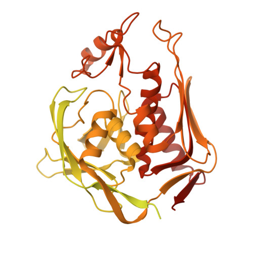
25RW | pdb_000025rw

Peptidoglycan and lipopolysaccharide biosynthesis enzymes with inhibitor


Experimental Data Snapshot

  • Method: ELECTRON MICROSCOPY
  • Resolution: 2.35 Å
  • Aggregation State: PARTICLE 
  • Reconstruction Method: SINGLE PARTICLE 

wwPDB Validation 3D Report Full Report

Validation slider image for 25RW

This is version 1.0 of the entry. See complete history. 

Literature

Structure of the MurA-LpxC enzyme complex in modulating peptidoglycan and lipopolysaccharide biosynthesis.

Yeo, J.Y., Yan, X.F., Qiao, Z., Liew, Y.Y., Do, P.H., Mu, Y., Gao, Y.G.

(2026) J Struct Biol : 108374-108374

  • DOI: https://doi.org/10.1016/j.jsb.2026.108374
  • Primary Citation Related Structures: 
    25RV, 25RW

  • PubMed Abstract: 

    Coordination of peptidoglycan and lipopolysaccharide biosynthesis is essential for maintaining Gram-negative cell envelope homeostasis. Two enzymes, MurA and LpxC, catalyze the first committed steps in peptidoglycan and lipopolysaccharide biosynthesis, respectively. Here, we determined cryo-electron microscopy (cryo-EM) structures of the Pseudomonas aeruginosa MurA-LpxC complex in the absence and presence of the LpxC inhibitor CHIR-090, providing molecular insights into complex formation. Structure-guided mutagenesis of MurA, together with in vitro pull-down assays, identified residues crucial for complex formation. We show that MurA G58 favors, but is not sufficient for complex formation, as substitution of this residue to mimic Escherichia coli MurA (G58S) weakens the interaction. Together, our study advances our structural understanding of how two biosynthesis pathways for peptidoglycan and lipopolysaccharide are coordinated to maintain a synergistic and balanced cell envelope.


  • Organizational Affiliation: 
    • School of Biological Sciences, Nanyang Technological University, 637551, Singapore; NTU Institute of Structural Biology, Nanyang Technological University, 636921, Singapore.

Macromolecule Content 

  • Total Structure Weight: 156.74 kDa 
  • Atom Count: 5,334 
  • Modeled Residue Count: 698 
  • Deposited Residue Count: 1,448 
  • Unique protein chains: 1

Macromolecules

Find similar proteins by:|  3D Structure
Entity ID: 1
MoleculeChains  Sequence LengthOrganismDetailsImage
UDP-N-acetylglucosamine 1-carboxyvinyltransferase,UDP-3-O-acyl-N-acetylglucosamine deacetylase
A, B
724Pseudomonas aeruginosa PAO1Mutation(s): 0 
Gene Names: murA, PA4450, lpxC, envA, PA4406
EC: 2.5.1.7 (PDB Primary Data), 3.5.1.108 (PDB Primary Data)
UniProt
Find proteins for Q9HVW7 (Pseudomonas aeruginosa (strain ATCC 15692 / DSM 22644 / CIP 104116 / JCM 14847 / LMG 12228 / 1C / PRS 101 / PAO1))
Explore Q9HVW7 
Go to UniProtKB:  Q9HVW7
Find proteins for P47205 (Pseudomonas aeruginosa (strain ATCC 15692 / DSM 22644 / CIP 104116 / JCM 14847 / LMG 12228 / 1C / PRS 101 / PAO1))
Explore P47205 
Go to UniProtKB:  P47205
Entity Groups
Sequence Clusters30% Identity50% Identity70% Identity90% Identity95% Identity100% Identity
UniProt GroupsP47205Q9HVW7
Sequence Annotations
Expand
Reference Sequence

Small Molecules

Ligands 1 Unique
IDChains Name / Formula / InChI Key2D Diagram3D Interactions
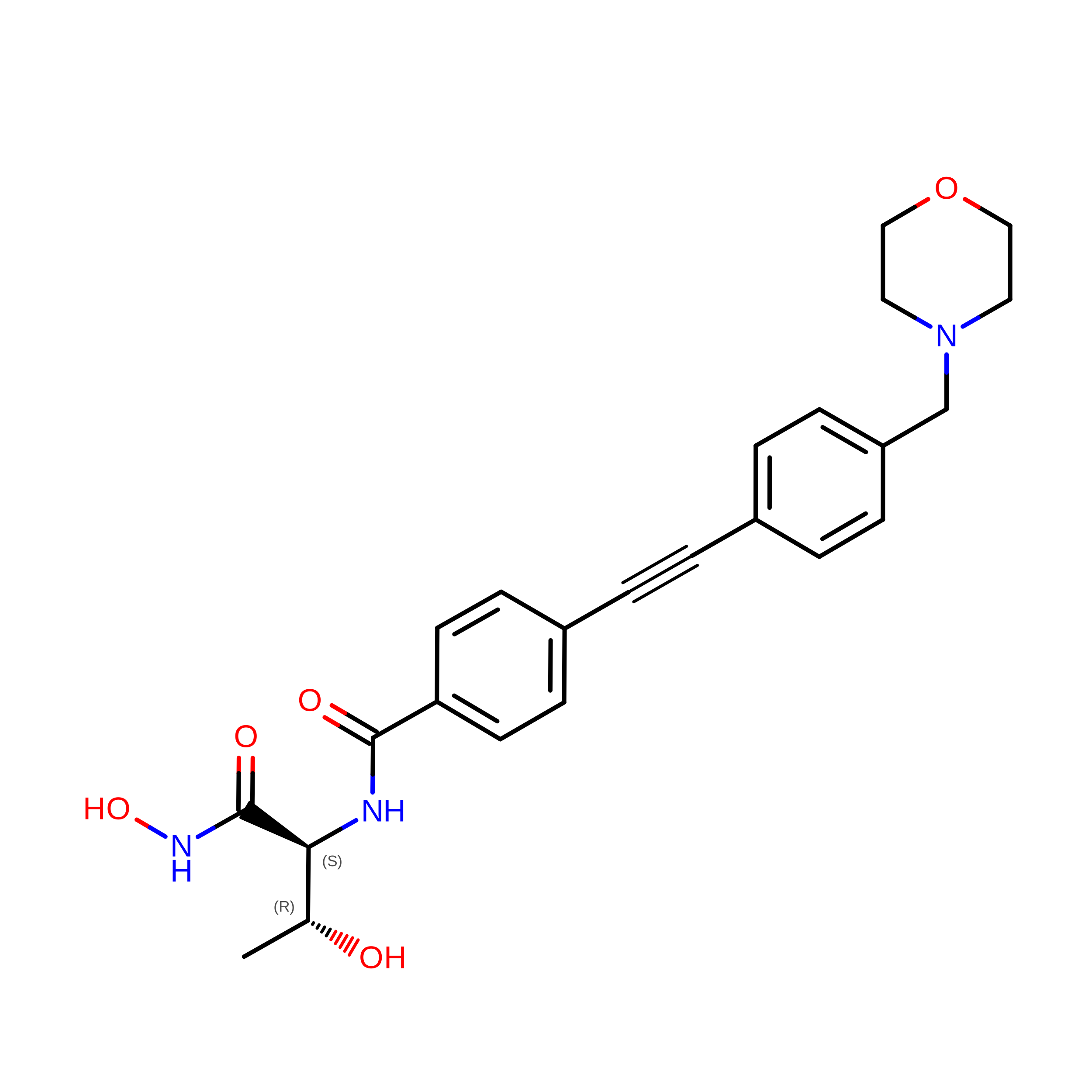
C90
(Subject of Investigation/LOI)

Query on C90


Download:Ideal Coordinates CCD File
C [auth A]N-{(1S,2R)-2-hydroxy-1-[(hydroxyamino)carbonyl]propyl}-4-{[4-(morpholin-4-ylmethyl)phenyl]ethynyl}benzamide
C24 H27 N3 O5
FQYBTYFKOHPWQT-VGSWGCGISA-N

Experimental Data & Validation

Experimental Data

  • Method: ELECTRON MICROSCOPY
  • Resolution: 2.35 Å
  • Aggregation State: PARTICLE 
  • Reconstruction Method: SINGLE PARTICLE 
EM Software:
TaskSoftware PackageVersion
MODEL REFINEMENTPHENIX2.0_5936
RECONSTRUCTIONcryoSPARC

Structure Validation

View Full Validation Report


Entry History 

& Funding Information

Deposition Data


Funding OrganizationLocationGrant Number
Ministry of Education (MoE, Singapore)Singapore--

Revision History  (Full details and data files)

  • Version 1.0: 2026-09-30
    Type: Initial release