23VY | pdb_000023vy

Crystal structure of the mouse RORalpha ligand binding domain in fusion with an NRIP1 LXXLL peptide


Experimental Data Snapshot

  • Method: X-RAY DIFFRACTION
  • Resolution: 2.70 Å
  • R-Value Free: 
    0.249 (Depositor), 0.248 (DCC) 
  • R-Value Work: 
    0.181 (Depositor), 0.181 (DCC) 
  • R-Value Observed: 
    0.188 (Depositor) 

Starting Model: experimental
View more details

wwPDB Validation   3D Report Full Report


Ligand Structure Quality Assessment 


This is version 1.1 of the entry. See complete history


Literature

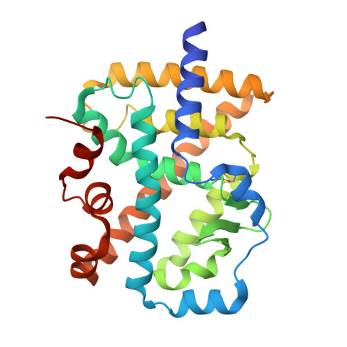
Structure of a chimeric ROR alpha ligand-binding domain in fusion with a RIP-140 coactivator peptide.

Li, X.Zhang, G.Tan, L.Chen, L.Im, Y.J.

(2026) Biochem Biophys Res Commun 812: 153622-153622

  • DOI: https://doi.org/10.1016/j.bbrc.2026.153622
  • Primary Citation Related Structures: 
    23VY

  • PubMed Abstract: 

    The retinoic acid-related orphan receptor α (RORα) is a potential drug target for cancer, inflammation, and metabolic diseases. Structure-guided ligand optimization can facilitate the development of selective RORα modulators. However, structural studies of the RORα ligand-binding domain (LBD) have been limited due to difficulties in purifying recombinant protein suitable for crystallographic analysis. Here, we engineered a chimeric RORα LBD C-terminally fused to the LXXLL motif of the coactivator RIP-140. This fusion improved the stability and solubility of the RORα LBD, enabling high-yield purification using the E. coli expression system. We determined the crystal structure of the chimeric RORα LBD in complex with cholesterol at 2.7 Å resolution. The cholesterol-bound RORα LBD adopted an active conformation of helix 12 that accommodates coactivator binding. The coactivator peptide appears to stabilize the RORα LBD by shielding the hydrophobic surface of the AF-2 region. Although monomeric in solution, the RORα LBD-LXXLL fusion formed a dimer in the crystal lattice through extensive interactions involving the LXXLL motifs and α3 helices, which may represent a physiological homodimer. Fusion of a coactivator motif to the LBD is expected to facilitate structural studies of RORα and may be broadly applicable to other nuclear receptor LBDs for generating stable recombinant proteins.


  • Organizational Affiliation
    • College of Pharmacy, Chonnam National University, Gwangju, 61186, Republic of Korea.

Macromolecules
Find similar proteins by:  (by identity cutoff)  |  3D Structure
Entity ID: 1
MoleculeChains Sequence LengthOrganismDetailsImage
Nuclear receptor ROR-alpha,undecapeptide
A, B
264Mus musculussynthetic construct
This entity is chimeric
Mutation(s): 0 
Gene Names: RoraNr1f1Rzra
UniProt & NIH Common Fund Data Resources
Find proteins for P51448 (Mus musculus)
Explore P51448 
Go to UniProtKB:  P51448
IMPC:  MGI:104661
Entity Groups  
Sequence Clusters30% Identity50% Identity70% Identity90% Identity95% Identity100% Identity
UniProt GroupP51448
Sequence Annotations
Expand
  • Reference Sequence
Experimental Data & Validation

Experimental Data

  • Method: X-RAY DIFFRACTION
  • Resolution: 2.70 Å
  • R-Value Free:  0.249 (Depositor), 0.248 (DCC) 
  • R-Value Work:  0.181 (Depositor), 0.181 (DCC) 
  • R-Value Observed: 0.188 (Depositor) 
Space Group: C 1 2 1
Unit Cell:
Length ( Å )Angle ( ˚ )
a = 115.804α = 90
b = 75.775β = 119.04
c = 81.661γ = 90
Software Package:
Software NamePurpose
HKL-2000data reduction
HKL-2000data scaling
PHASERphasing
Cootmodel building
PHENIXrefinement

Structure Validation

View Full Validation Report


Ligand Structure Quality Assessment 


Entry History & Funding Information

Deposition Data


Funding OrganizationLocationGrant Number
National Research Foundation (NRF, Korea)Korea, Republic OfRS-2023-00221356

Revision History  (Full details and data files)

  • Version 1.0: 2026-03-11
    Type: Initial release
  • Version 1.1: 2026-04-01
    Changes: Database references