23ON | pdb_000023on

Crystal Structure Analysis of Bovine Carbonic Anhydrase II to 4-2-methyl-1,3-oxazol-5-ylbenzenesulfonamide


Experimental Data Snapshot

  • Method: X-RAY DIFFRACTION
  • Resolution: 1.50 Å
  • R-Value Free: 
    0.200 (Depositor), 0.200 (DCC) 
  • R-Value Work: 
    0.155 (Depositor), 0.156 (DCC) 
  • R-Value Observed: 
    0.158 (Depositor) 

Starting Model: experimental
View more details

wwPDB Validation   3D Report Full Report


Ligand Structure Quality Assessment 


This is version 1.1 of the entry. See complete history


Literature

X-ray analysis of complexes of carbonic anhydrase II with 1,3-oxazole-containing sulfonamide derivatives elucidates the structural basis for their exceptionally high inhibitory potency.

Stoliarskaia, M.Y.Borisevich, S.S.Myaskov, E.O.Shetnev, A.A.Korsakov, M.K.Nikonov, O.S.

(2026) Acta Crystallogr F Struct Biol Commun 

  • DOI: https://doi.org/10.1107/S2053230X26002128
  • Primary Citation Related Structures: 
    23OM, 23ON

  • PubMed Abstract: 

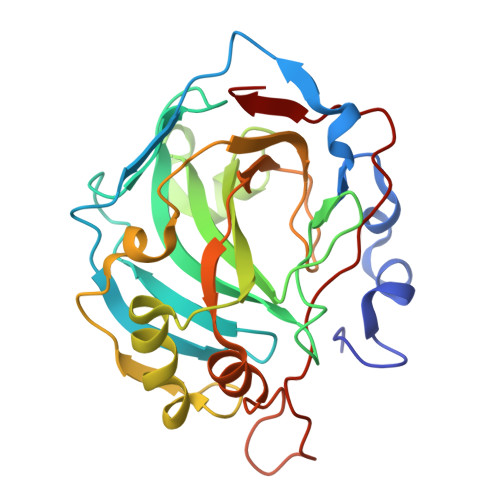
    Carbonic anhydrase II (CA2) is a zinc metalloenzyme that catalyzes the reversible hydration of CO 2 and is widely used as a model for structure-guided inhibitor development. Previously reported crystal structures of human CA II (α-hCA2) in complex with the picomolar sulfonamide inhibitors 8V5 and 8V8 (PDB entries 5nee and 5nea) did not allow the complete ligand geometry of 8V5 to be resolved. To clarify the structural basis of binding, we determined high-resolution (1.5-1.7 Å) crystal structures of bovine CA II (α-bCA2) in complex with both inhibitors. α-bCA2 was selected as a crystallographically robust homolog, and structural alignment with α-hCA2 showed an r.m.s.d. of <0.44 Å across C α atoms, confirming a near-identical active-site architecture. The α-bCA2-8V8 complex reproduced the canonical sulfonamide-zinc coordination pattern. In contrast, the α-bCA2-8V5 structure revealed continuous electron density for the entire ligand, including the morpholine group, which was not observed previously. Re-refinement of the α-hCA2-8V5 model based on these data resulted in an improved fit to density, indicating that the absence of the morpholine density in the original model is consistent with partial ligand modification or variability in the ligand state during crystallization. These results demonstrate that cross-isoform structural comparison can resolve ambiguities in ligand modeling and provide a reliable framework for the rational design of selective CA2 inhibitors.


  • Organizational Affiliation
    • Institute of Protein Research, Institutskaya 4, Pushchino, Moscow Region, Russian Federation.

Macromolecules
Find similar proteins by:  (by identity cutoff)  |  3D Structure
Entity ID: 1
MoleculeChains Sequence LengthOrganismDetailsImage
Carbonic anhydrase 2
A, B
259Bos taurusMutation(s): 0 
Gene Names: CA2
EC: 4.2.1.1 (PDB Primary Data), 4.2.1.69 (PDB Primary Data)
UniProt
Find proteins for P00921 (Bos taurus)
Explore P00921 
Go to UniProtKB:  P00921
Entity Groups  
Sequence Clusters30% Identity50% Identity70% Identity90% Identity95% Identity100% Identity
UniProt GroupP00921
Sequence Annotations
Expand
  • Reference Sequence
Small Molecules
Ligands 4 Unique
IDChains Name / Formula / InChI Key2D Diagram3D Interactions
8V8 (Subject of Investigation/LOI)
Query on 8V8

Download Ideal Coordinates CCD File 
C [auth A],
J [auth B]
4-(2-methyl-1,3-oxazol-5-yl)benzenesulfonamide
C10 H10 N2 O3 S
FETRXICILYMBJH-UHFFFAOYSA-N
GOL
Query on GOL

Download Ideal Coordinates CCD File 
D [auth A]
E [auth A]
G [auth A]
I [auth B]
K [auth B]
D [auth A],
E [auth A],
G [auth A],
I [auth B],
K [auth B],
L [auth B]
GLYCEROL
C3 H8 O3
PEDCQBHIVMGVHV-UHFFFAOYSA-N
ZN (Subject of Investigation/LOI)
Query on ZN

Download Ideal Coordinates CCD File 
H [auth A],
O [auth B]
ZINC ION
Zn
PTFCDOFLOPIGGS-UHFFFAOYSA-N
ACT
Query on ACT

Download Ideal Coordinates CCD File 
F [auth A],
M [auth B],
N [auth B]
ACETATE ION
C2 H3 O2
QTBSBXVTEAMEQO-UHFFFAOYSA-M
Experimental Data & Validation

Experimental Data

  • Method: X-RAY DIFFRACTION
  • Resolution: 1.50 Å
  • R-Value Free:  0.200 (Depositor), 0.200 (DCC) 
  • R-Value Work:  0.155 (Depositor), 0.156 (DCC) 
  • R-Value Observed: 0.158 (Depositor) 
Space Group: P 1 21 1
Unit Cell:
Length ( Å )Angle ( ˚ )
a = 59.773α = 90
b = 80.163β = 106.94
c = 61.771γ = 90
Software Package:
Software NamePurpose
PHENIXrefinement
XDSdata reduction
XDSdata scaling
MOLREPphasing

Structure Validation

View Full Validation Report


Ligand Structure Quality Assessment 


Entry History & Funding Information

Deposition Data


Funding OrganizationLocationGrant Number
Not fundedRussian Federation--

Revision History  (Full details and data files)

  • Version 1.0: 2026-02-25
    Type: Initial release
  • Version 1.1: 2026-03-25
    Changes: Database references