23FH | pdb_000023fh

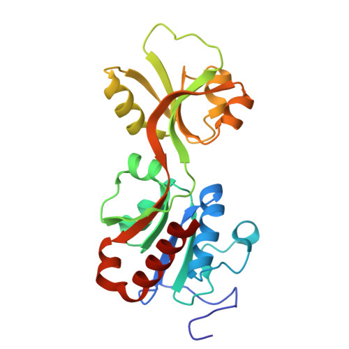
Crystal structure of short-form adenosine triphosphate phosphoribosyltransferase from Acinetobacter baumannii at 2.18 angstrom resolution.


Experimental Data Snapshot

  • Method: X-RAY DIFFRACTION
  • Resolution: 2.18 Å
  • R-Value Free: 
    0.212 (Depositor), 0.218 (DCC) 
  • R-Value Work: 
    0.179 (Depositor), 0.191 (DCC) 

Starting Model: experimental
View more details

wwPDB Validation 3D Report Full Report

Validation slider image for 23FH

This is version 1.1 of the entry. See complete history

Literature

Structure of the short-form ATP-phosphoribosyltransferase from Acinetobacter baumannii reveals the presence of a novel Mg 2+ sequestering and storage site.

Ahmad, N.Sharma, P.Sharma, S.Singh, T.P.

(2026) Biochim Biophys Acta Proteins Proteom 1874: 141159-141159

  • DOI: https://doi.org/10.1016/j.bbapap.2026.141159
  • Primary Citation Related Structures: 
    23FH, 23FI, 7WGM, 7WZY

  • PubMed Abstract: 

    Adenosine triphosphate phosphoribosyltransferase (ATP-PRT), a member of the phosphoribosyltransferase (PRT) superfamily of enzymes, catalyses the first step in the histidine biosynthesis pathway, which involves the nucleophilic substitution of ATP onto phosphoribosyl pyrophosphate (PRPP) to generate phosphoribosyl-ATP (PR-ATP) and pyrophosphate (PP i ). The three-dimensional structure of short-form ATP-PRT from Acinetobacter baumannii (AbHisG S ) was determined using the X-ray crystallographic method. The structure determination revealed two crystallographically independent molecules in the asymmetric unit that formed an antiparallel homodimer. In all the structures of HisG S proteins reported so far, the N-terminal segment is either absent from the amino acid sequence or not observed in the structure. This is the first structure where the N-terminal segment has been clearly observed. However, the most remarkable observation pertains to observing a Mg 2+ ion in the structure with the help of the N-terminal segment, which is loosely held with six coordination linkages, out of which one contact is made to the carbonyl oxygen atom of Asn16 at an average distance of 2.06 Å, and the other five contacts were with water oxygen atoms at distances varying from 2.40 to 2.59 Å. All the coordinating water oxygen atoms were held in place by multiple hydrogen bonds from surrounding protein atoms. The relatively loose coordination environment of the Mg 2+ ion indicates temporary sequestration of Mg 2+ , which may be released during catalytic need.


  • Organizational Affiliation
    • Department of Biophysics, All India Institute of Medical Sciences, New Delhi 110029, India.

Macromolecule Content 

  • Total Structure Weight: 50.58 kDa 
  • Atom Count: 3,808 
  • Modeled Residue Count: 452 
  • Deposited Residue Count: 454 
  • Unique protein chains: 1

Macromolecules

Find similar proteins by:|  3D Structure
Entity ID: 1
MoleculeChains  Sequence LengthOrganismDetailsImage
ATP phosphoribosyltransferase
A, B
227Acinetobacter baumanniiMutation(s): 0 
EC: 2.4.2.17
UniProt
Find proteins for A0ABF7PKC5 (Acinetobacter baumannii)
Explore A0ABF7PKC5 
Go to UniProtKB:  A0ABF7PKC5
Entity Groups
Sequence Clusters30% Identity50% Identity70% Identity90% Identity95% Identity100% Identity
UniProt GroupA0ABF7PKC5
Sequence Annotations
Expand
Reference Sequence

Small Molecules

Ligands 3 Unique
IDChains Name / Formula / InChI Key2D Diagram3D Interactions
ACT

Query on ACT


Download:Ideal Coordinates CCD File
C [auth A],
D [auth A]
ACETATE ION
C2 H3 O2
QTBSBXVTEAMEQO-UHFFFAOYSA-M
FMT

Query on FMT


Download:Ideal Coordinates CCD File
E [auth A],
F [auth A],
G [auth A],
I [auth B]
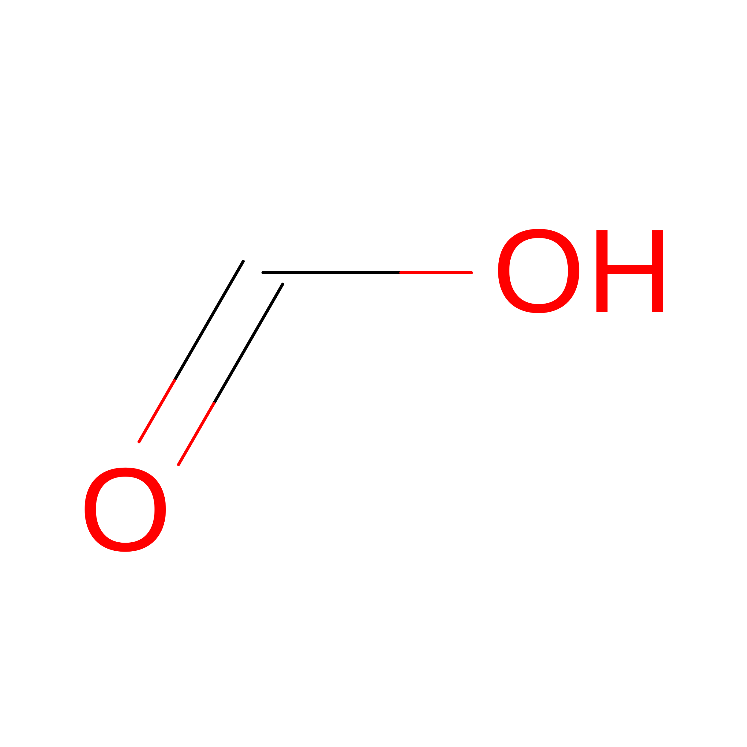
FORMIC ACID
C H2 O2
BDAGIHXWWSANSR-UHFFFAOYSA-N
MG
(Subject of Investigation/LOI)

Query on MG


Download:Ideal Coordinates CCD File
H [auth A],
J [auth B]
MAGNESIUM ION
Mg
JLVVSXFLKOJNIY-UHFFFAOYSA-N

Experimental Data & Validation

Experimental Data

  • Method: X-RAY DIFFRACTION
  • Resolution: 2.18 Å
  • R-Value Free:  0.212 (Depositor), 0.218 (DCC) 
  • R-Value Work:  0.179 (Depositor), 0.191 (DCC) 
Space Group: P 21 21 21
Unit Cell:
Length ( Å )Angle ( ˚ )
a = 71.476α = 90
b = 79.203β = 90
c = 97.777γ = 90
Software Package:
Software NamePurpose
REFMACrefinement
XDSdata reduction
Aimlessdata scaling
MOLREPphasing
MxCuBEdata collection
Cootmodel building

Structure Validation

View Full Validation Report


Entry History 

& Funding Information

Deposition Data


Funding OrganizationLocationGrant Number
Indian Council of Medical ResearchIndiaI-1252

Revision History  (Full details and data files)

  • Version 1.0: 2026-02-18
    Type: Initial release
  • Version 1.1: 2026-07-15
    Changes: Database references