22VH | pdb_000022vh

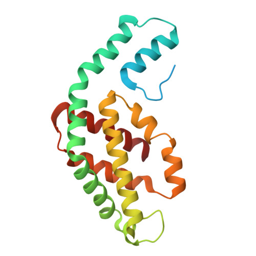
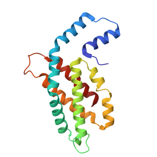
The assembled CPC trimer


Experimental Data Snapshot

  • Method: X-RAY DIFFRACTION
  • Resolution: 2.50 Å
  • R-Value Free: 
    0.246 (Depositor), 0.246 (DCC) 
  • R-Value Work: 
    0.194 (Depositor), 0.194 (DCC) 
  • R-Value Observed: 
    0.196 (Depositor) 

Starting Model: experimental
View more details

wwPDB Validation 3D Report Full Report

Validation slider image for 22VH

Ligand Structure Quality Assessment 


This is version 1.0 of the entry. See complete history

Literature

Biosynthesis of C-phycocyanin trimers.

Zhao, X.Zhu, J.X.Li, G.C.Zhao, K.H.

(2026) Biochim Biophys Acta Bioenerg 1867: 149599-149599

  • DOI: https://doi.org/10.1016/j.bbabio.2026.149599
  • Primary Citation Related Structures: 
    22VH

  • PubMed Abstract: 

    Phycobiliproteins, particularly C-phycocyanin (CPC), serve as major light-harvesting complexes in cyanobacteria, exhibiting high efficiency in light-harvesting and energy transfer. Currently, the easy and large-scale acquisition of functional CPC remains a major challenge, primarily due to the requirement for precise, sequential covalent attachment of multiple phycocyanobilin (PCB) chromophores. In this study, by using a dual-promoter (T7 and araBAD) system to control the sequential binding of two PCB chromophores to β82 and β153, we successfully achieved the biosynthesis of β-CPC (λ max, absorption  = 605 nm, λ max, emission  = 644 nm) in E. coli, which can transfer energy from the β153-PCB to the β82-PCB. The assembly of α-CPC with PCB-β82, PCB-β153, and PCB 2 -β under identical conditions indicates that only PCB 2 -β, which covalently binds the two PCB chromophores, can assemble with α-CPC to form a complete CPC trimer (λ max, absorption  = 617 nm, λ max, emission  = 646 nm). The structure of the assembled trimer reveals that it adopts a typical phycobiliprotein fold, with multiple chromophores precisely arranged, exhibiting features highly similar to those of native CPC. Furthermore, we established a biosynthetic pathway for CPC trimers in E. coli. This system provides a powerful tool for engineering phycobiliproteins with various light-harvesting and energy transfer properties, facilitating future studies on artificial photosynthesis, light-harvesting antenna design, and the fundamental mechanisms of excitation energy transfer.


  • Organizational Affiliation
    • National Key Laboratory of Agricultural Microbiology, Huazhong Agricultural University, Wuhan, 430070, PR China.

Macromolecule Content 

  • Total Structure Weight: 250.18 kDa 
  • Atom Count: 16,584 
  • Modeled Residue Count: 2,028 
  • Deposited Residue Count: 2,232 
  • Unique protein chains: 2

Macromolecules

Find similar proteins by:|  3D Structure
Entity ID: 1
MoleculeChains  Sequence LengthOrganismDetailsImage
C-phycocyanin alpha subunit
A,
C [auth B],
E,
G,
I,
K
199Nostoc sp. PCC 7120 = FACHB-418Mutation(s): 0 
Gene Names: cpcAalr0529
UniProt
Find proteins for P07121 (Nostoc sp. (strain PCC 7120 / SAG 25.82 / UTEX 2576))
Explore P07121 
Go to UniProtKB:  P07121
Entity Groups
Sequence Clusters30% Identity50% Identity70% Identity90% Identity95% Identity100% Identity
UniProt GroupP07121
Sequence Annotations
Expand
Reference Sequence
Find similar proteins by:|  3D Structure
Entity ID: 2
MoleculeChains  Sequence LengthOrganismDetailsImage
C-phycocyanin beta subunit173Nostoc sp. PCC 7120 = FACHB-418Mutation(s): 0 
Gene Names: cpcBalr0528
UniProt
Find proteins for P07120 (Nostoc sp. (strain PCC 7120 / SAG 25.82 / UTEX 2576))
Explore P07120 
Go to UniProtKB:  P07120
Entity Groups
Sequence Clusters30% Identity50% Identity70% Identity90% Identity95% Identity100% Identity
UniProt GroupP07120
Sequence Annotations
Expand
Reference Sequence

Small Molecules

Ligands 1 Unique
IDChains Name / Formula / InChI Key2D Diagram3D Interactions
CYC
(Subject of Investigation/LOI)

Query on CYC


Download:Ideal Coordinates CCD File
AA [auth J]
BA [auth K]
CA [auth L]
DA [auth L]
M [auth A]
AA [auth J],
BA [auth K],
CA [auth L],
DA [auth L],
M [auth A],
N [auth D],
O [auth D],
P [auth B],
Q [auth C],
R [auth C],
S [auth E],
T [auth F],
U [auth F],
V [auth G],
W [auth H],
X [auth H],
Y [auth I],
Z [auth J]
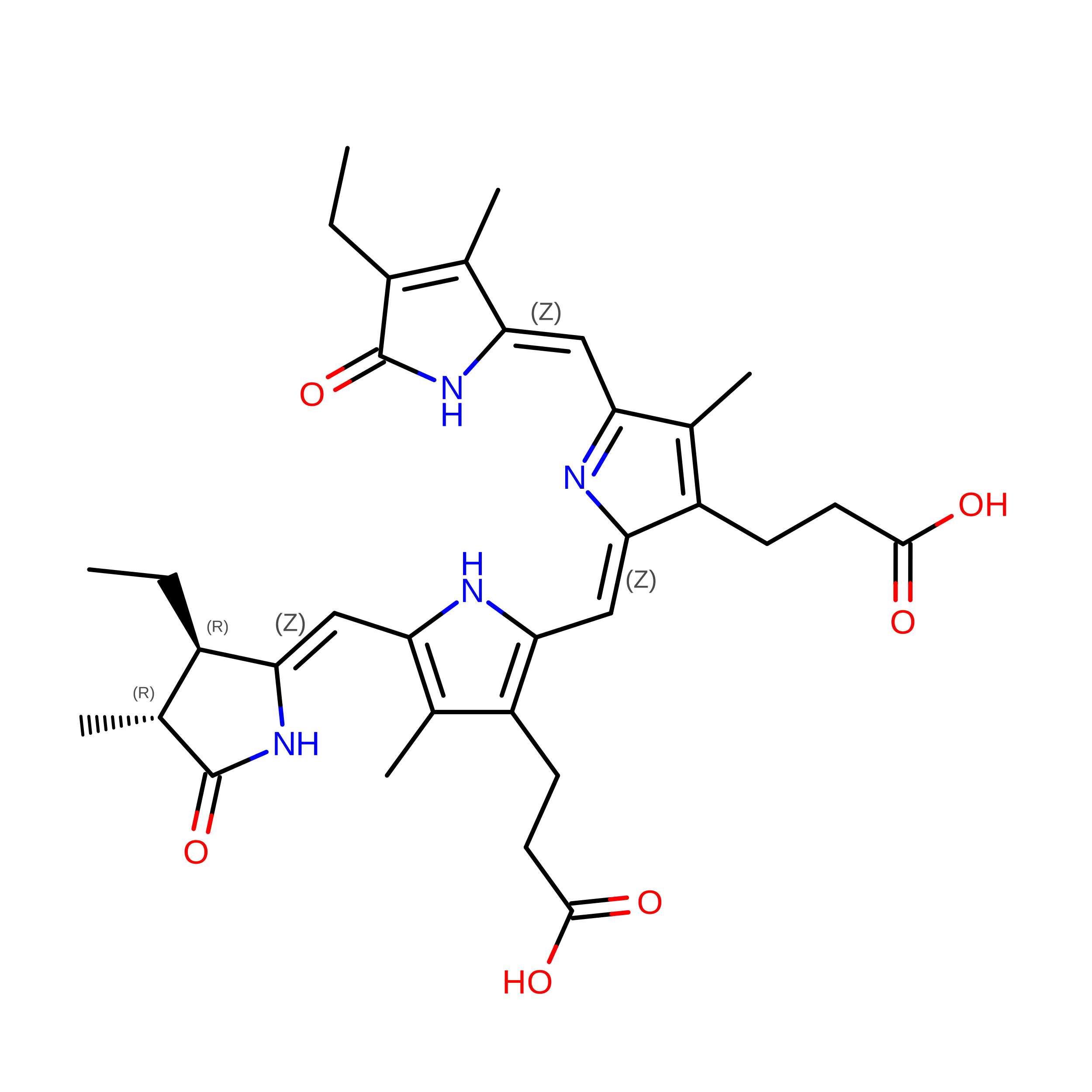
PHYCOCYANOBILIN
C33 H40 N4 O6
VXTXPYZGDQPMHK-GMXXPEQVSA-N

Experimental Data & Validation

Experimental Data

  • Method: X-RAY DIFFRACTION
  • Resolution: 2.50 Å
  • R-Value Free:  0.246 (Depositor), 0.246 (DCC) 
  • R-Value Work:  0.194 (Depositor), 0.194 (DCC) 
  • R-Value Observed: 0.196 (Depositor) 
Space Group: C 1 2 1
Unit Cell:
Length ( Å )Angle ( ˚ )
a = 117.97α = 90
b = 109.96β = 103.47
c = 182.37γ = 90
Software Package:
Software NamePurpose
PHENIXrefinement
XDSdata reduction
PHASERphasing
Aimlessdata scaling

Structure Validation

View Full Validation Report


Ligand Structure Quality Assessment 


Entry History 

& Funding Information

Deposition Data


Funding OrganizationLocationGrant Number
National Natural Science Foundation of China (NSFC)China31861143029

Revision History  (Full details and data files)

  • Version 1.0: 2026-08-19
    Type: Initial release