Download Mendeley
1 H, 13 C, and 15 N resonance assignments and solution structure of the first double-stranded RNA-binding domain of DExH-Box Helicase 30 (DHX30).
Abe, C., Muto, Y., He, F., Takahashi, M., Tsuda, K., Nagata, T., Tanaka, A., Kobayashi, N., Kigawa, T., Guntert, P., Shirouzu, M., Yokoyama, S., Kuwasako, K.(2026) Biomol NMR Assign 20
- PubMed: 42850484 Search on PubMed
- DOI: https://doi.org/10.1007/s12104-026-10279-0
- Primary Citation Related Structures:
22KA - PubMed Abstract:
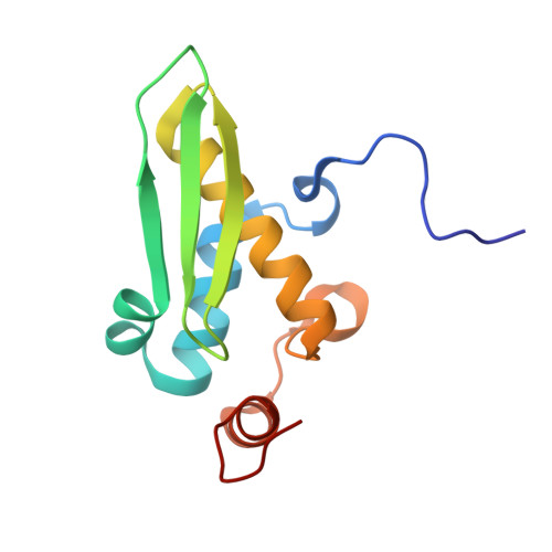
DHX30 is a member of the DExH-box helicase family and contains two double-stranded RNA-binding domains (dsRBDs) N-terminal to the helicase core domain. DHX30 plays a fundamental role in regulating the translation of transcripts encoding mitoribosomal proteins, thereby facilitating mitoribosome biogenesis. Additionally, DHX30 is involved in the formation of stress granules. These intracellular structures are composed of proteins and RNAs, including mRNAs, and form under stress conditions in association with translational repression. Recent studies have demonstrated that pathogenic mutations in DHX30 underlie a severe neurodevelopmental disorder termed neurodevelopmental disorder with variable motor and language impairment (NEDMIAL), possibly in part through aberrant stress-granule formation. Structural characterization of these RNA-binding domains is an important step toward understanding the molecular basis of DHX30 function. In this study, we focus on the first dsRBD (dsRBD-1) of human DHX30 and report its 1 H, 13 C, and 15 N resonance assignments and solution structure. DHX30 dsRBD-1 adopts an α0-α1-α1'-β1-β2-β3-α2-α3 topology, with three additional helices (α0, α1', and α3) relative to the canonical dsRBD topology (α1-β1-β2-β3-α2). To our knowledge, this is the first experimentally determined structure of a DHX30 domain. These resonance assignments and structural insights provide a valuable foundation for future structural and functional studies of DHX30.
- RIKEN, Systems and Structural Biology Center, 1-7-22 Suehiro-cho, Tsurumi-ku, Yokohama, 230-0045, Japan.
Organizational Affiliation: