22JW | pdb_000022jw

Perinereis linea erythrocruorin


Experimental Data Snapshot

  • Method: ELECTRON MICROSCOPY
  • Resolution: 2.84 Å
  • Aggregation State: PARTICLE 
  • Reconstruction Method: SINGLE PARTICLE 

wwPDB Validation 3D Report Full Report

Validation slider image for 22JW

This is version 1.0 of the entry. See complete history

Literature

Structure of Perinereis linea erythrocruorin reveals a compact extracellular globin megacomplex.

Deng, J.X.Cheng, W.B.Xu, K.Hou, P.Chen, Y.Jiang, Y.L.Zhou, C.Z.

(2026) Structure 

  • DOI: https://doi.org/10.1016/j.str.2026.06.004
  • Primary Citation Related Structures: 
    22JW

  • PubMed Abstract: 

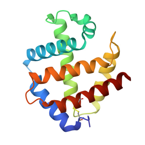
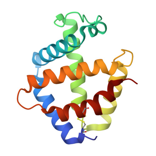
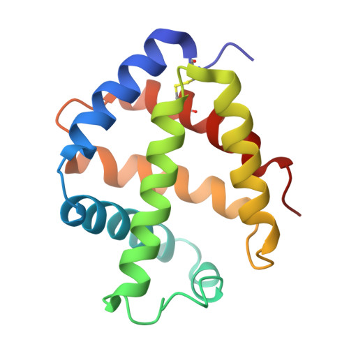
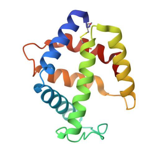
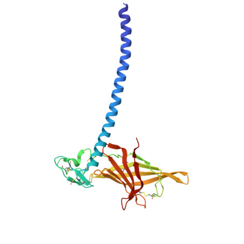
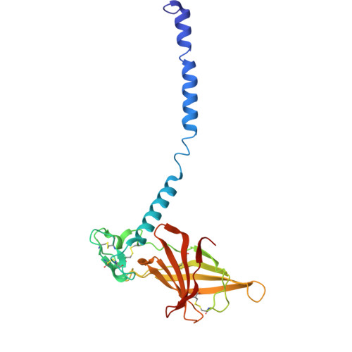
    Many invertebrates lack erythrocytes and instead rely on extracellular hemoglobin assemblies, termed erythrocruorins, for oxygen transport. Here we report a 2.84 Å cryo-electron microscopy (cryo-EM) structure of Perinereis linea erythrocruorin (PlEc). PlEc is a ∼3.3 MDa megacomplex composed of 180 polypeptide chains organized into 12 protomers, forming a hexagonal bilayer with D6 symmetry. Each protomer consists of 12 globin subunits and three linker subunits, adopting a mushroom-like architecture. The cap of the mushroom is formed by a globin dodecamer associated with a heterotrimeric linker head, and the stem consists of a triple-stranded coiled coil derived from the N-terminal helices of three linker subunits. Biochemical assays show that PlEc has thermal stability and auto-oxidation rate comparable to those of other erythrocruorins, but displays relatively lower oxygen-binding affinity. These findings provide mechanistic insights into the quaternary assembly of invertebrate erythrocruorins and lay the groundwork for the potential biomedical applications.


  • Organizational Affiliation
    • Department of Radiology, the First Affiliated Hospital of USTC, and School of Life Sciences, Division of Life Sciences and Medicine, University of Science and Technology of China, Hefei 230026, China.

Macromolecule Content 

  • Total Structure Weight: 279.37 kDa 
  • Atom Count: 19,644 
  • Modeled Residue Count: 2,408 
  • Deposited Residue Count: 2,408 
  • Unique protein chains: 6

Macromolecules

Find similar proteins by:|  3D Structure
Entity ID: 1
MoleculeChains  Sequence LengthOrganismDetailsImage
Extracellular globin A
A, E, I
148Perinereis lineaMutation(s): 0 
Entity Groups
Sequence Clusters30% Identity50% Identity70% Identity90% Identity95% Identity100% Identity
Sequence Annotations
Expand
Reference Sequence
Find similar proteins by:|  3D Structure
Entity ID: 2
MoleculeChains  Sequence LengthOrganismDetailsImage
Extracellular globin B
B, F, J
145Perinereis lineaMutation(s): 0 
Entity Groups
Sequence Clusters30% Identity50% Identity70% Identity90% Identity95% Identity100% Identity
Sequence Annotations
Expand
Reference Sequence
Find similar proteins by:|  3D Structure
Entity ID: 3
MoleculeChains  Sequence LengthOrganismDetailsImage
Extracellular globin C
C, G, K
148Perinereis lineaMutation(s): 0 
Entity Groups
Sequence Clusters30% Identity50% Identity70% Identity90% Identity95% Identity100% Identity
Sequence Annotations
Expand
Reference Sequence
Find similar proteins by:|  3D Structure
Entity ID: 4
MoleculeChains  Sequence LengthOrganismDetailsImage
Extracellular globin D
D, H, L
138Perinereis lineaMutation(s): 0 
Entity Groups
Sequence Clusters30% Identity50% Identity70% Identity90% Identity95% Identity100% Identity
Sequence Annotations
Expand
Reference Sequence
Find similar proteins by:|  3D Structure
Entity ID: 5
MoleculeChains  Sequence LengthOrganismDetailsImage
Hemoglobin linker 1
M, N
223Perinereis lineaMutation(s): 0 
Entity Groups
Sequence Clusters30% Identity50% Identity70% Identity90% Identity95% Identity100% Identity
Sequence Annotations
Expand
Reference Sequence
Find similar proteins by:|  3D Structure
Entity ID: 6
MoleculeChains  Sequence LengthOrganismDetailsImage
Hemoglobin linker 2225Perinereis lineaMutation(s): 0 
Entity Groups
Sequence Clusters30% Identity50% Identity70% Identity90% Identity95% Identity100% Identity
Sequence Annotations
Expand
Reference Sequence

Small Molecules

Ligands 2 Unique
IDChains Name / Formula / InChI Key2D Diagram3D Interactions
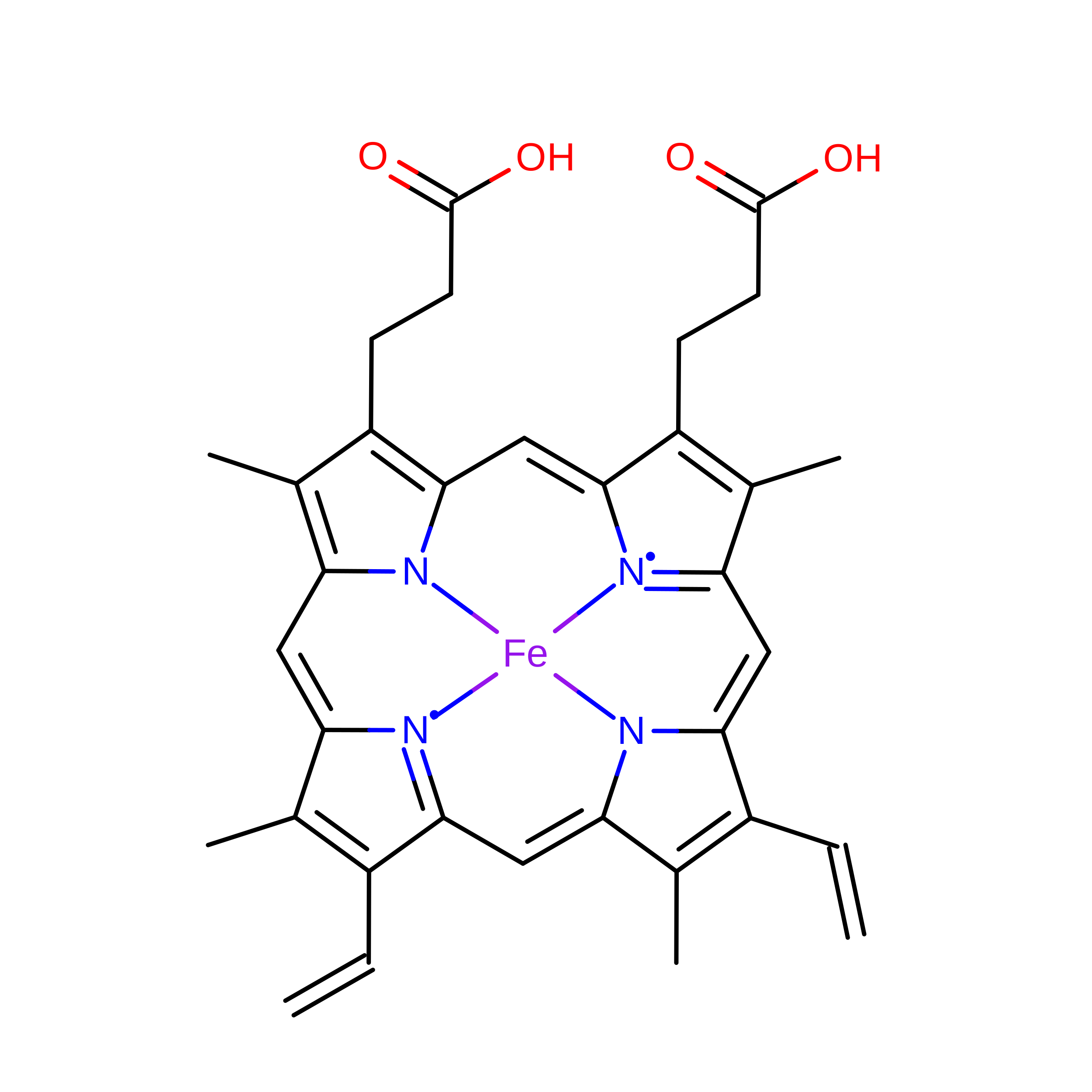
HEM
(Subject of Investigation/LOI)

Query on HEM


Download:Ideal Coordinates CCD File
AA [auth L]
P [auth A]
Q [auth B]
R [auth C]
S [auth D]
AA [auth L],
P [auth A],
Q [auth B],
R [auth C],
S [auth D],
T [auth E],
U [auth F],
V [auth G],
W [auth H],
X [auth I],
Y [auth J],
Z [auth K]
PROTOPORPHYRIN IX CONTAINING FE
C34 H32 Fe N4 O4
KABFMIBPWCXCRK-RGGAHWMASA-L
CA
(Subject of Investigation/LOI)

Query on CA


Download:Ideal Coordinates CCD File
BA [auth M],
CA [auth N],
DA [auth O]
CALCIUM ION
Ca
BHPQYMZQTOCNFJ-UHFFFAOYSA-N

Experimental Data & Validation

Experimental Data

  • Method: ELECTRON MICROSCOPY
  • Resolution: 2.84 Å
  • Aggregation State: PARTICLE 
  • Reconstruction Method: SINGLE PARTICLE 
EM Software:
TaskSoftware PackageVersion
MODEL REFINEMENTPHENIX1.20.1_4487
RECONSTRUCTIONcryoSPARC

Structure Validation

View Full Validation Report


Entry History 

& Funding Information

Deposition Data


Funding OrganizationLocationGrant Number
National Natural Science Foundation of China (NSFC)China32241025

Revision History  (Full details and data files)

  • Version 1.0: 2026-08-12
    Type: Initial release