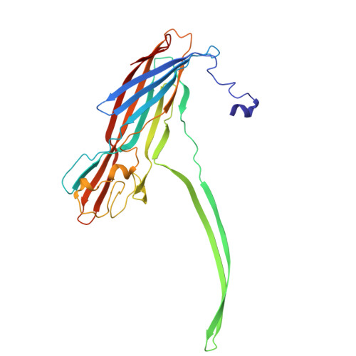
21GF | pdb_000021gf

Structure of the heptameric pore state of alpha-hemolysin in the presence of A549 cells at pH 5.5


Experimental Data Snapshot

  • Method: ELECTRON MICROSCOPY
  • Resolution: 3.75 Å
  • Aggregation State: PARTICLE 
  • Reconstruction Method: SINGLE PARTICLE 

wwPDB Validation 3D Report Full Report

Validation slider image for 21GF

This is version 1.0 of the entry. See complete history

Literature

Structure of the heptameric pore state of alpha-hemolysin in the presence of A549 cells at pH 5.5

Chatterjee, A.Dutta, S.

To be published.

Macromolecule Content 

  • Total Structure Weight: 233.03 kDa 
  • Atom Count: 16,415 
  • Modeled Residue Count: 2,051 
  • Deposited Residue Count: 2,051 
  • Unique protein chains: 1

Macromolecules

Find similar proteins by:|  3D Structure
Entity ID: 1
MoleculeChains  Sequence LengthOrganismDetailsImage
Alpha-hemolysin
A, B, C, D, E
A, B, C, D, E, F, G
293Staphylococcus aureusMutation(s): 0 
Gene Names: hlyhla
UniProt
Find proteins for P09616 (Staphylococcus aureus)
Explore P09616 
Go to UniProtKB:  P09616
Entity Groups
Sequence Clusters30% Identity50% Identity70% Identity90% Identity95% Identity100% Identity
UniProt GroupP09616
Sequence Annotations
Expand
Reference Sequence

Experimental Data & Validation

Experimental Data

  • Method: ELECTRON MICROSCOPY
  • Resolution: 3.75 Å
  • Aggregation State: PARTICLE 
  • Reconstruction Method: SINGLE PARTICLE 
EM Software:
TaskSoftware PackageVersion
RECONSTRUCTIONcryoSPARC

Structure Validation

View Full Validation Report


Entry History 

& Funding Information

Deposition Data


Funding OrganizationLocationGrant Number
Department of Biotechnology (DBT, India)India--

Revision History  (Full details and data files)

  • Version 1.0: 2026-08-05
    Type: Initial release