Download Mendeley
Cryo-EM structure of the native assembled Mfa type V pilus from the periodontal pathogen Porphyromonas gingivalis.
Shibata, S., Matsunami, H., Ouhara, K., Taniguchi, Y., Schreiber, M.T., Villar-Brillones, A., Nakayama, K., Shoji, M., Wolf, M.(2026) Commun Biol
- PubMed: 42336997 Search on PubMed
- DOI: https://doi.org/10.1038/s42003-026-10515-2
- Primary Citation Related Structures:
21CO - PubMed Abstract:
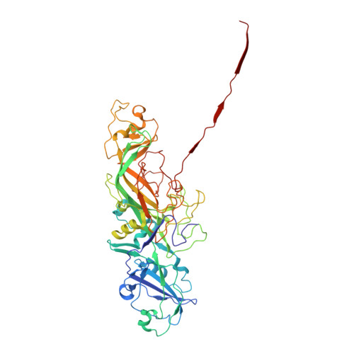
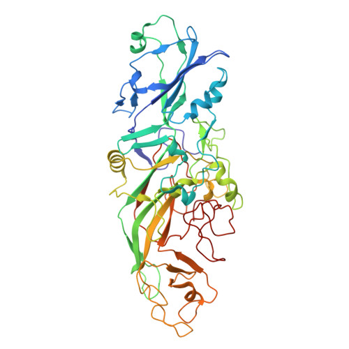
Porphyromonas gingivalis is a primary pathogen causing periodontal disease. The cell has two kinds of type V pili, the Fim pilus and the Mfa pilus, both of which play essential roles in colonization, biofilm formation, and pathogenicity. The functional polymerized structure of the Fim pilus is known, whereas the structure and assembly mechanism of the Mfa pilus remain unclear. Here, we show the structure of the polymerized recombinant Mfa1 stalk pilin determined by cryo-electron microscopy at 3.0 Å resolution. The atomic model of the Mfa1 filament reveals that Mfa1 pilins polymerize by protease-mediated strand exchange and retain a Ca 2+ ion in the metal-binding pocket, which modulates immune recognition of the Mfa pilus by human cells. Furthermore, we elucidated the three-dimensional architecture of the streptococcal-binding region on the Mfa pilus. Our results further strengthen evidence that protease-mediated strand exchange is the universal assembly mechanism of type V pili. Our structure of the polymerized Mfa pilus, which represents the functional state on the cell surface, provides targets for antimicrobial drug design to treat periodontal disease and P. gingivalis-related systemic diseases.
- Molecular Cryo-Electron Microscopy Unit, Okinawa Institute of Science and Technology Graduate University, Okinawa, Japan. sshibata@tottori-u.ac.jp.
Organizational Affiliation: