 Download Mendeley
Cryo-EM structure of ex vivo Sup35 yeast prion and the factors defining prion phenotype
Dergalev, A.A., Chesnokov, Y.M., Burtseva, A.D., Mitkevich, O.V., Baymukhametov, T.N., Popov, V.O., Boyko, K.M., Kushnirov, V.V.(2026) Nat Commun
Experimental Data Snapshot
Starting Model: other
View more details
wwPDB Validation 3D Report Full Report

(2026) Nat Commun
Entity ID: 1 | |||||
|---|---|---|---|---|---|
| Molecule | Chains | Sequence Length | Organism | Details | Image |
| Amyloid of yeast prion Sup35 | 160 | Saccharomyces cerevisiae | Mutation(s): 0 | 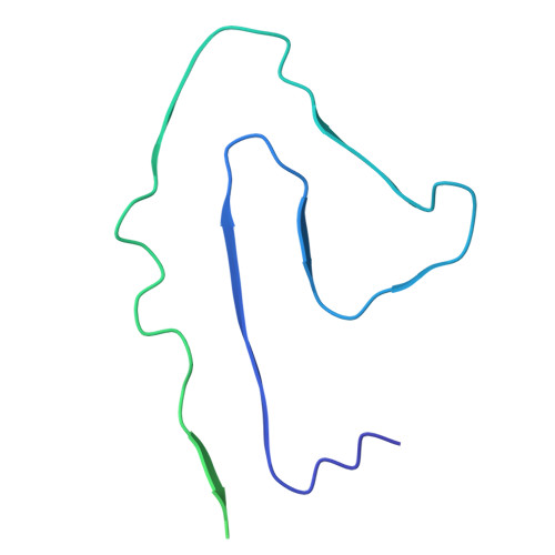 | |
Entity Groups | |||||
| Sequence Clusters | 30% Identity50% Identity70% Identity90% Identity95% Identity100% Identity | ||||
Sequence AnnotationsExpand | |||||
Reference Sequence | |||||
| Task | Software Package | Version |
|---|---|---|
| RECONSTRUCTION | cryoSPARC | 4.6 |
| MODEL REFINEMENT | PHENIX | 1.21.2_5419 |
| Funding Organization | Location | Grant Number |
|---|---|---|
| Russian Science Foundation | Russian Federation | 23-74-00062 |
