20ZX | pdb_000020zx

Cryo-EM structure of Gq-coupled LPAR5 in complex with LPA


Experimental Data Snapshot

  • Method: ELECTRON MICROSCOPY
  • Resolution: 2.96 Å
  • Aggregation State: PARTICLE 
  • Reconstruction Method: SINGLE PARTICLE 

wwPDB Validation 3D Report Full Report

Validation slider image for 20ZX

This is version 1.2 of the entry. See complete history

Literature

Structural basis for lysophosphatidic acid recognition and atypical G alpha q coupling by LPAR5.

Li, X.Wang, K.Xing, Z.Zhang, M.Hu, W.Yuan, Q.Xu, H.E.Zhao, L.H.

(2026) Proc Natl Acad Sci U S A 123

  • DOI: https://doi.org/10.1073/pnas.2537482123
  • Primary Citation Related Structures: 
    20ZX

  • PubMed Abstract: 

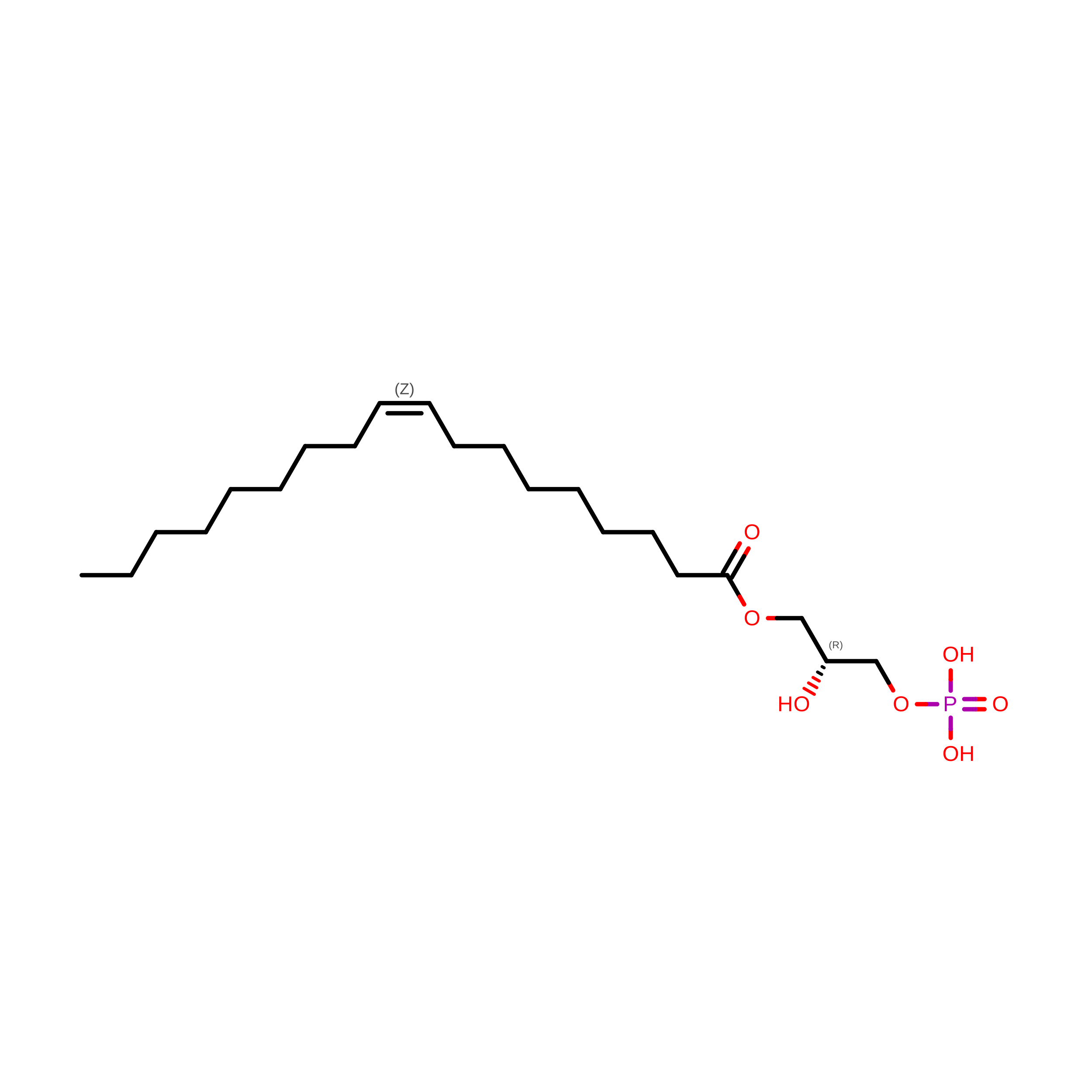
    Lysophosphatidic acid receptor 5 (LPAR5) is a non-endothelial differentiation gene class A G protein-coupled receptor that regulates neuropathic pain, itch, and cancer progression through coupling to G proteins. Here, we report the cryo-EM structure of LPAR5 bound to 1-oleoyl-lysophosphatidic acid (LPA) in complex with G q at 2.96 Å resolution, revealing a distinct mode of receptor activation and G protein coupling. The phosphate headgroup of LPA forms extensive polar interactions with residues from extracellular loop 2 and transmembrane helices TM5-TM7, while the lipid tail inserts into a deep hydrophobic cavity formed by TM3-TM5. Site-directed mutagenesis confirms the functional importance of these interactions. Remarkably, LPAR5 exhibits a noncanonical G protein coupling mode. Unlike previously reported GPCR-G protein structures in which the Gα C-terminal α5 helix ("wavy hook") primarily engages TM6, the wavy hook in LPAR5 is positioned toward the intracellular loop 1-helix 8 interface. This configuration is associated with limited TM6 outward displacement and modest rearrangement at the toggle-switch position (6.48). The resulting interface is stabilized by receptor-specific interactions and supported by functional data. Together, these findings reveal an alternative mode of GPCR-G protein coupling and highlight the structural plasticity underlying signaling specificity in LPA receptors.


  • Organizational Affiliation
    • State Key Laboratory of Drug Research, Shanghai Institute of Materia Medica, Chinese Academy of Sciences, Shanghai 201203, China.

Macromolecule Content 

  • Total Structure Weight: 157.78 kDa 
  • Atom Count: 9,077 
  • Modeled Residue Count: 1,149 
  • Deposited Residue Count: 1,421 
  • Unique protein chains: 5

Macromolecules

Find similar proteins by:|  3D Structure
Entity ID: 1
MoleculeChains  Sequence LengthOrganismDetailsImage
Guanine nucleotide-binding protein G(I)/G(S)/G(T) subunit beta-1A [auth B]371Rattus norvegicusMutation(s): 0 
Gene Names: Gnb1
UniProt
Find proteins for P54311 (Rattus norvegicus)
Explore P54311 
Go to UniProtKB:  P54311
Entity Groups
Sequence Clusters30% Identity50% Identity70% Identity90% Identity95% Identity100% Identity
UniProt GroupP54311
Sequence Annotations
Expand
Reference Sequence
Find similar proteins by:|  3D Structure
Entity ID: 2
MoleculeChains  Sequence LengthOrganismDetailsImage
Guanine nucleotide-binding protein G(I)/G(S)/G(O) subunit gamma-2B [auth G]70Bos taurusMutation(s): 0 
Gene Names: GNG2
UniProt
Find proteins for P63212 (Bos taurus)
Explore P63212 
Go to UniProtKB:  P63212
Entity Groups
Sequence Clusters30% Identity50% Identity70% Identity90% Identity95% Identity100% Identity
UniProt GroupP63212
Sequence Annotations
Expand
Reference Sequence
Find similar proteins by:|  3D Structure
Entity ID: 3
MoleculeChains  Sequence LengthOrganismDetailsImage
Lysophosphatidic acid receptor 5C [auth R]372Homo sapiensMutation(s): 0 
Gene Names: LPAR5GPR92GPR93
UniProt & NIH Common Fund Data Resources
Find proteins for Q9H1C0 (Homo sapiens)
Explore Q9H1C0 
Go to UniProtKB:  Q9H1C0
GTEx:  ENSG00000184574 
Entity Groups
Sequence Clusters30% Identity50% Identity70% Identity90% Identity95% Identity100% Identity
UniProt GroupQ9H1C0
Sequence Annotations
Expand
Reference Sequence
Find similar proteins by:|  3D Structure
Entity ID: 4
MoleculeChains  Sequence LengthOrganismDetailsImage
Guanine nucleotide-binding protein G(324) subunit alphaD [auth A]361Homo sapiensMutation(s): 0 
Entity Groups
Sequence Clusters30% Identity50% Identity70% Identity90% Identity95% Identity100% Identity
Sequence Annotations
Expand
Reference Sequence
Find similar proteins by:|  3D Structure
Entity ID: 5
MoleculeChains  Sequence LengthOrganismDetailsImage
scFv16247Homo sapiensMutation(s): 0 
Entity Groups
Sequence Clusters30% Identity50% Identity70% Identity90% Identity95% Identity100% Identity
Sequence Annotations
Expand
Reference Sequence

Small Molecules

Ligands 1 Unique
IDChains Name / Formula / InChI Key2D Diagram3D Interactions
UBL
(Subject of Investigation/LOI)

Query on UBL


Download:Ideal Coordinates CCD File
F [auth R][(2R)-2-oxidanyl-3-phosphonooxy-propyl] (Z)-octadec-9-enoate
C21 H41 O7 P
WRGQSWVCFNIUNZ-GDCKJWNLSA-N

Experimental Data & Validation

Experimental Data

  • Method: ELECTRON MICROSCOPY
  • Resolution: 2.96 Å
  • Aggregation State: PARTICLE 
  • Reconstruction Method: SINGLE PARTICLE 
EM Software:
TaskSoftware PackageVersion
MODEL REFINEMENTPHENIX
RECONSTRUCTIONcryoSPARC

Structure Validation

View Full Validation Report


Entry History 

& Funding Information

Deposition Data


Funding OrganizationLocationGrant Number
National Natural Science Foundation of China (NSFC)China32371255
National Natural Science Foundation of China (NSFC)China32130022

Revision History  (Full details and data files)

  • Version 1.0: 2026-06-17
    Type: Initial release
  • Version 1.1: 2026-06-24
    Changes: Data collection, Database references, Source and taxonomy, Structure summary
  • Version 1.2: 2026-07-01
    Changes: Data collection, Database references