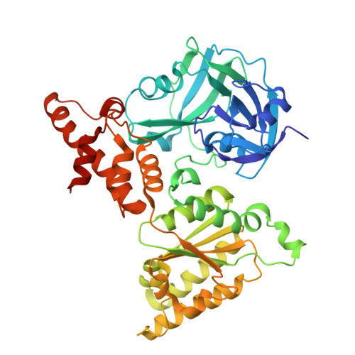
20ZO | pdb_000020zo

XFEL structure of p97 N-D1 hexamer mimicking ATP-bound conformation

  • Classification: HYDROLASE
  • Organism(s): Homo sapiens
  • Expression System: Escherichia coli
  • Mutation(s): No 

  • Deposited: 2025-12-03 Released: 2026-10-28 
  • Deposition Author(s): Kang, W.
  • Funding Organization(s): National Research Foundation (NRF, Korea)

Experimental Data Snapshot

  • Method: X-RAY DIFFRACTION
  • Resolution: 2.00 Å
  • R-Value Free: 
    0.254 (Depositor), 0.251 (DCC) 
  • R-Value Work: 
    0.218 (Depositor), 0.215 (DCC) 
  • R-Value Observed: 
    0.220 (Depositor) 

Starting Model: experimental
View more details

wwPDB Validation 3D Report Full Report

Validation slider image for 20ZO

Ligand Structure Quality Assessment 


This is version 1.0 of the entry. See complete history. 

Literature

Structural insights into the ADP-bound p97/VCP ND1 hexamer revealed by room-temperature XFEL crystallography.

Koh, S., Jeon, H., Lee, E., Kim, Y., Jang, S., Cho, H., Shin, M., Cho, S., Choi, R., Jung, S., Jung, S., Ham, Y., Kim, Y., Park, J., Park, S., Park, S., Lee, S.J., Yang, J.K., Kang, W.

(2026) Int J Biol Macromol 380: 154062-154062

  • DOI: https://doi.org/10.1016/j.ijbiomac.2026.154062
  • Primary Citation Related Structures: 
    20ZO, 20ZP

  • PubMed Abstract: 

    p97/VCP ATPase uses adenosine triphosphate (ATP) hydrolysis to remodel protein assemblies and unfold substrates across diverse cellular processes, including protein quality control, membrane fusion, and chromatin regulation. Although cryogenic structural studies have provided valuable snapshots of distinct nucleotide states, they often suppress the conformational variations that are intrinsic to the protein function. Here, we present a comparative structural analysis of the ADP-bound human p97 ND1 hexamer using room-temperature X-ray free-electron laser (XFEL) and cryogenic synchrotron crystallography. While the fundamental hexameric architecture is conserved across both conditions, the room-temperature XFEL structure reveals significantly enhanced conformational variability. Notably, the N-terminal domain exhibits increased flexibility and positional variations. Furthermore, the room-temperature structure reveals an extended, ordered active-site water network that connects the nucleotide-binding pocket to the bulk solvent, a feature absent under cryogenic conditions. Interestingly, the ADP ribose undergoes a pucker transition from a canonical C2'endo to a C3'-endo conformation, revealing altered active-site conformations. Together, these structures highlight the intrinsic conformational flexibility of the p97 ND1 fragment using room-temperature XFEL crystallography as a powerful approach for identifying temperature-dependent structural flexibility of ATPases.


  • Organizational Affiliation: 
    • Department of Chemistry, College of Natural Sciences, Soongsil University, Seoul, 06978, Republic of Korea.

Macromolecule Content 

  • Total Structure Weight: 50.31 kDa 
  • Atom Count: 3,540 
  • Modeled Residue Count: 437 
  • Deposited Residue Count: 445 
  • Unique protein chains: 1

Macromolecules

Find similar proteins by:|  3D Structure
Entity ID: 1
MoleculeChains  Sequence LengthOrganismDetailsImage
Transitional endoplasmic reticulum ATPase445Homo sapiensMutation(s): 0 
Gene Names: VCP, HEL-220, HEL-S-70
EC: 3.6.4.6
UniProt & NIH Common Fund Data Resources
Find proteins for P55072 (Homo sapiens)
Explore P55072 
Go to UniProtKB:  P55072
PHAROS:  P55072
GTEx:  ENSG00000165280 
Entity Groups
Sequence Clusters30% Identity50% Identity70% Identity90% Identity95% Identity100% Identity
UniProt GroupP55072
Sequence Annotations
Expand
Reference Sequence

Small Molecules

Ligands 1 Unique
IDChains Name / Formula / InChI Key2D Diagram3D Interactions
ADP
(Subject of Investigation/LOI)

Query on ADP


Download:Ideal Coordinates CCD File
B [auth A]ADENOSINE-5'-DIPHOSPHATE
C10 H15 N5 O10 P2
XTWYTFMLZFPYCI-KQYNXXCUSA-N

Experimental Data & Validation

Experimental Data

  • Method: X-RAY DIFFRACTION
  • Resolution: 2.00 Å
  • R-Value Free:  0.254 (Depositor), 0.251 (DCC) 
  • R-Value Work:  0.218 (Depositor), 0.215 (DCC) 
  • R-Value Observed: 0.220 (Depositor) 
Space Group: P 6 2 2
Unit Cell:
Length ( Å )Angle ( ˚ )
a = 145.98α = 90
b = 145.98β = 90
c = 86.53γ = 120
Software Package:
Software NamePurpose
PHENIXrefinement
Cheetahdata collection
CrystFELdata reduction
CrystFELdata scaling
PHASERphasing

Structure Validation

View Full Validation Report


Ligand Structure Quality Assessment 


Entry History 

& Funding Information

Deposition Data

  • Released Date: 2026-10-28 
  • Deposition Author(s): Kang, W.

Funding OrganizationLocationGrant Number
National Research Foundation (NRF, Korea)Korea, Republic OfRS-2022-NR071729
National Research Foundation (NRF, Korea)Korea, Republic OfRS-2021-NR060140
National Research Foundation (NRF, Korea)Korea, Republic OfRS-2023-00217189
National Research Foundation (NRF, Korea)Korea, Republic OfRS-2025-25441317

Revision History  (Full details and data files)

  • Version 1.0: 2026-10-28
    Type: Initial release