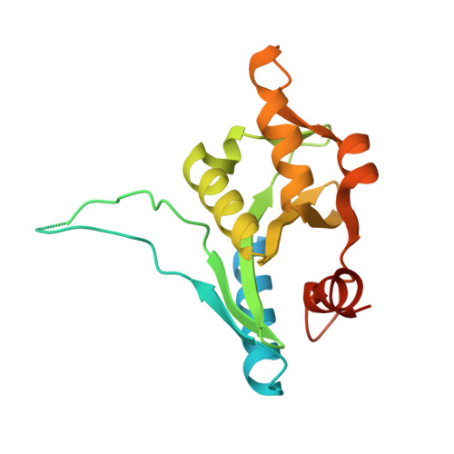
1ZP7

The structure of Bacillus subtilis RecU Holliday junction resolvase and its role in substrate selection and sequence specific cleavage.


Experimental Data Snapshot

  • Method: X-RAY DIFFRACTION
  • Resolution: 2.25 Å
  • R-Value Free: 0.269 
  • R-Value Work: 0.217 
  • R-Value Observed: 0.219 

wwPDB Validation   3D Report Full Report


This is version 1.3 of the entry. See complete history


Literature

The Structure of Bacillus subtilis RecU Holliday Junction Resolvase and Its Role in Substrate Selection and Sequence-Specific Cleavage.

McGregor, N.Ayora, S.Sedelnikova, S.Carrasco, B.Alonso, J.C.Thaw, P.Rafferty, J.

(2005) Structure 13: 1341-1351

  • DOI: https://doi.org/10.1016/j.str.2005.05.011
  • Primary Citation of Related Structures:  
    1ZP7

  • PubMed Abstract: 

    We have determined the structure of the enzyme RecU from Bacillus subtilis, that is the general Holliday junction resolving enzyme in Gram-positive bacteria. The enzyme fold reveals a striking similarity to a class of resolvase enzymes found in archaeal sources and members of the type II restriction endonuclease family to which they are related. The structure confirms the presence of active sites formed around clusters of acidic residues that we have also shown to bind divalent cations. Mutagenesis data presented here support the key role of certain residues. The RecU structure suggests a basis for Holliday junction selectivity and suggests how sequence-specific cleavage might be achieved. Models for a resolvase-DNA complex address how the enzyme might organize junctions into an approximately 4-fold symmetric form.


  • Organizational Affiliation

    The Krebs Institute, Department of Molecular Biology and Biotechnology, University of Sheffield, Western Bank, Sheffield S10 2TN, United Kingdom.


Macromolecules
Find similar proteins by:  (by identity cutoff)  |  3D Structure
Entity ID: 1
MoleculeChains Sequence LengthOrganismDetailsImage
Recombination protein U
A, B
206Bacillus subtilisMutation(s): 0 
EC: 3.1.21.10
UniProt
Find proteins for P39792 (Bacillus subtilis (strain 168))
Explore P39792 
Go to UniProtKB:  P39792
Entity Groups  
Sequence Clusters30% Identity50% Identity70% Identity90% Identity95% Identity100% Identity
UniProt GroupP39792
Sequence Annotations
Expand
  • Reference Sequence
Experimental Data & Validation

Experimental Data

  • Method: X-RAY DIFFRACTION
  • Resolution: 2.25 Å
  • R-Value Free: 0.269 
  • R-Value Work: 0.217 
  • R-Value Observed: 0.219 
  • Space Group: P 65
Unit Cell:
Length ( Å )Angle ( ˚ )
a = 74.34α = 90
b = 74.34β = 90
c = 153.466γ = 120
Software Package:
Software NamePurpose
REFMACrefinement
MOSFLMdata reduction
CCP4data scaling
SOLVEphasing

Structure Validation

View Full Validation Report


Entry History 

Deposition Data

Revision History  (Full details and data files)

  • Version 1.0: 2005-09-20
    Type: Initial release
  • Version 1.1: 2008-04-30
    Changes: Version format compliance
  • Version 1.2: 2011-07-13
    Changes: Version format compliance
  • Version 1.3: 2024-02-14
    Changes: Data collection, Database references