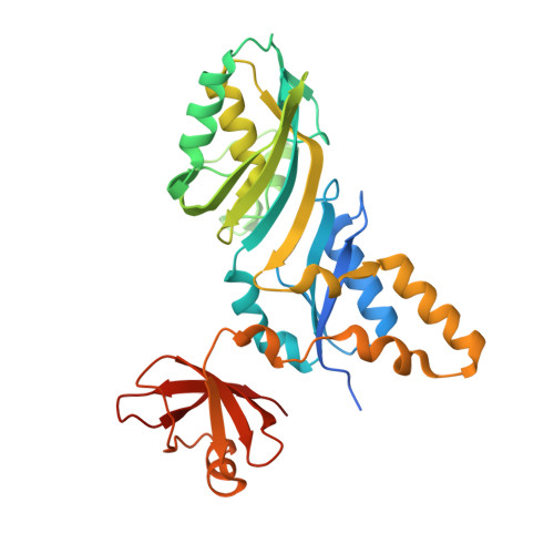
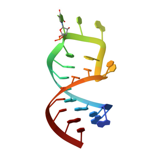
1ZL3

Coupling of active site motions and RNA binding


Experimental Data Snapshot

  • Method: X-RAY DIFFRACTION
  • Resolution: 2.80 Å
  • R-Value Free: 0.264 
  • R-Value Work: 0.202 
  • R-Value Observed: 0.202 

Starting Model: experimental
View more details

wwPDB Validation   3D Report Full Report


This is version 1.4 of the entry. See complete history


Literature

Precursor complex structure of pseudouridine synthase TruB suggests coupling of active site perturbations to an RNA-sequestering peripheral protein domain

Hoang, C.Hamilton, C.S.Mueller, E.G.Ferre-D'Amare, A.R.

(2005) Protein Sci 14: 2201-2206

  • DOI: https://doi.org/10.1110/ps.051493605
  • Primary Citation of Related Structures:  
    1ZL3

  • PubMed Abstract: 

    The pseudouridine synthase TruB is responsible for the universally conserved post-transcriptional modification of residue 55 of elongator tRNAs. In addition to the active site, the "thumb", a peripheral domain unique to the TruB family of enzymes, makes extensive interactions with the substrate. To coordinate RNA binding and release with catalysis, the thumb may be able to sense progress of the reaction in the active site. To establish whether there is a structural correlate of communication between the active site and the RNA-sequestering thumb, we have solved the structure of a catalytically inactive point mutant of TruB in complex with a substrate RNA, and compared it to the previously determined structure of an active TruB bound to a reaction product. Superposition of the two structures shows that they are extremely similar, except in the active site and, intriguingly, in the relative position of the thumb. Because the two structures were solved using isomorphous crystals, and because the thumb is very well ordered in both structures, the displacement of the thumb we observe likely reflects preferential propagation of active site perturbations to this RNA-binding domain. One of the interactions between the active site and the thumb involves an active site residue whose hydrogen-bonding status changes during the reaction. This may allow the peripheral RNA-binding domain to monitor progress of the pseudouridylation reaction.


  • Organizational Affiliation

    Division of Basic Sciences, Fred Hutchinson Cancer Research Center, Seattle, WA 98109-1024, USA.


Macromolecules

Find similar proteins by:  (by identity cutoff)  |  3D Structure
Entity ID: 2
MoleculeChains Sequence LengthOrganismDetailsImage
tRNA pseudouridine synthase BB [auth A]327Escherichia coliMutation(s): 1 
Gene Names: truB
EC: 4.2.1.70 (PDB Primary Data), 5.4.99.25 (UniProt)
UniProt
Find proteins for P60340 (Escherichia coli (strain K12))
Explore P60340 
Go to UniProtKB:  P60340
Entity Groups  
Sequence Clusters30% Identity50% Identity70% Identity90% Identity95% Identity100% Identity
UniProt GroupP60340
Sequence Annotations
Expand
  • Reference Sequence

Find similar nucleic acids by:  Sequence   |   3D Structure  

Entity ID: 1
MoleculeChains LengthOrganismImage
5'-R(*GP*GP*CP*AP*AP*CP*GP*GP*UP*(FLO) UP*CP*GP*AP*UP*CP*CP*CP*GP*UP*UP*GP*C)-3'A [auth B]22N/A
Sequence Annotations
Expand
  • Reference Sequence
Experimental Data & Validation

Experimental Data

  • Method: X-RAY DIFFRACTION
  • Resolution: 2.80 Å
  • R-Value Free: 0.264 
  • R-Value Work: 0.202 
  • R-Value Observed: 0.202 
  • Space Group: C 1 2 1
Unit Cell:
Length ( Å )Angle ( ˚ )
a = 145.433α = 90
b = 40.753β = 110.61
c = 78.803γ = 90
Software Package:
Software NamePurpose
DENZOdata reduction
SCALEPACKdata scaling
CNSrefinement
CNSphasing

Structure Validation

View Full Validation Report


Entry History 

Deposition Data

Revision History  (Full details and data files)

  • Version 1.0: 2005-08-09
    Type: Initial release
  • Version 1.1: 2008-04-30
    Changes: Version format compliance
  • Version 1.2: 2011-07-13
    Changes: Version format compliance
  • Version 1.3: 2021-10-20
    Changes: Database references, Derived calculations
  • Version 1.4: 2023-08-23
    Changes: Data collection, Refinement description