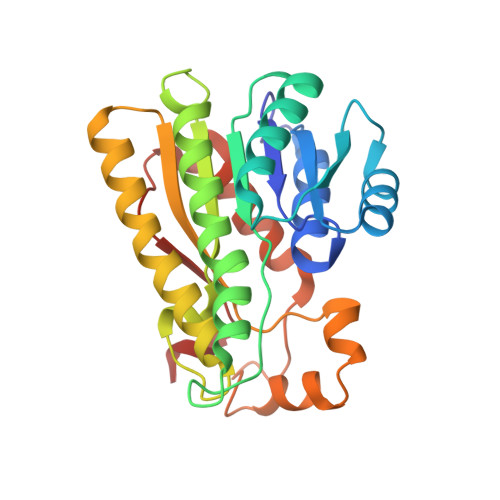
Atomic Resolution Structures of R-specific Alcohol Dehydrogenase from Lactobacillus brevis Provide the Structural Bases of its Substrate and Cosubstrate Specificity
Schlieben, N.H., Niefind, K., Muller, J., Riebel, B., Hummel, W., Schomburg, D.(2005) J Mol Biol 349: 801-813
- PubMed: 15896805 Search on PubMed
- DOI: https://doi.org/10.1016/j.jmb.2005.04.029
- Primary Citation Related Structures:
1ZJY, 1ZJZ, 1ZK0, 1ZK1, 1ZK2, 1ZK3, 1ZK4 - PubMed Abstract:
The R-specific alcohol dehydrogenase (RADH) from Lactobacillus brevis is an NADP-dependent, homotetrameric member of the extended enzyme family of short-chain dehydrogenases/reductases (SDR) with a high biotechnological application potential. Its preferred in vitro substrates are prochiral ketones like acetophenone with almost invariably a small methyl group as one substituent and a bulky (often aromatic) moiety as the other. On the basis of an atomic-resolution structure of wild-type RADH in complex with NADP and acetophenone, we designed the mutant RADH-G37D, which should possess an improved cosubstrate specificity profile for biotechnological purposes, namely, a preference for NAD rather than NADP. Comparative kinetic measurements with wild-type and mutant RADH showed that this aim was achieved. To characterize the successful mutant structurally, we determined several, partly atomic-resolution, crystal structures of RADH-G37D both as an apo-enzyme and as ternary complex with NAD or NADH and phenylethanol. The increased affinity of RADH-G37D for NAD(H) depends on an interaction between the adenosine ribose moiety of NAD and the inserted aspartate side-chain. A structural comparison between RADH-G37D as apo-enzyme and as a part of a ternary complex revealed significant rearrangements of Ser141, Glu144, Tyr189 and Met205 in the vicinity of the active site. This plasticity contributes to generate a small hydrophobic pocket for the methyl group typical for RADH substrates, and a hydrophobic coat for the second, more variable and often aromatic, substituent. Around Ser141 we even found alternative conformations in the backbone. A structural adaptability in this region, which we describe here for the first time for an SDR enzyme, is probably functionally important, because it concerns Ser142, a member of the highly conserved catalytic tetrad typical for SDR enzymes. Moreover, it affects an extended proton relay system that has been identified recently as a critical element for the catalytic mechanism in SDR enzymes.
- Universität zu Köln, Institut für Biochemie, Zülpicher Strasse 47, D-50674 Köln, Germany.
Organizational Affiliation: