1ZID | pdb_00001zid

LONG FATTY ACID CHAIN ENOYL-ACP REDUCTASE (INHA) IN COMPLEX WITH AN ISONICOTINIC-ACYL-NADH INHIBITOR


Experimental Data Snapshot

  • Method: X-RAY DIFFRACTION
  • Resolution: 2.70 Å
  • R-Value Free: 
    0.297 (Depositor), 0.287 (DCC) 
  • R-Value Work: 
    0.202 (Depositor), 0.197 (DCC) 
  • R-Value Observed: 
    0.202 (Depositor) 

Starting Model: experimental
View more details

wwPDB Validation 3D Report Full Report

Validation slider image for 1ZID

Ligand Structure Quality Assessment 


This is version 1.4 of the entry. See complete history

Literature

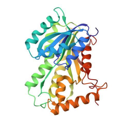
Modification of the NADH of the isoniazid target (InhA) from Mycobacterium tuberculosis.

Rozwarski, D.A.Grant, G.A.Barton, D.H.Jacobs Jr., W.R.Sacchettini, J.C.

(1998) Science 279: 98-102

  • DOI: https://doi.org/10.1126/science.279.5347.98
  • Primary Citation Related Structures: 
    1ZID

  • PubMed Abstract: 

    The preferred antitubercular drug isoniazid specifically targets a long-chain enoyl-acyl carrier protein reductase (InhA), an enzyme essential for mycolic acid biosynthesis in Mycobacterium tuberculosis. Despite the widespread use of this drug for more than 40 years, its precise mode of action has remained obscure. Data from x-ray crystallography and mass spectrometry reveal that the mechanism of isoniazid action against InhA is covalent attachment of the activated form of the drug to the nicotinamide ring of nicotinamide adenine dinucleotide bound within the active site of InhA.


  • Organizational Affiliation
    • Department of Biochemistry and Biophysics, Texas A&M University, College Station, TX 77843, USA.

Macromolecule Content 

  • Total Structure Weight: 29.16 kDa 
  • Atom Count: 2,114 
  • Modeled Residue Count: 268 
  • Deposited Residue Count: 268 
  • Unique protein chains: 1

Macromolecules

Find similar proteins by:|  3D Structure
Entity ID: 1
MoleculeChains  Sequence LengthOrganismDetailsImage
ENOYL-[ACYL-CARRIER-PROTEIN] REDUCTASE268Mycobacterium tuberculosisMutation(s): 0 
EC: 1.3.1.9
UniProt
Find proteins for P9WGR1 (Mycobacterium tuberculosis (strain ATCC 25618 / H37Rv))
Explore P9WGR1 
Go to UniProtKB:  P9WGR1
Entity Groups
Sequence Clusters30% Identity50% Identity70% Identity90% Identity95% Identity100% Identity
UniProt GroupP9WGR1
Sequence Annotations
Expand
Reference Sequence

Small Molecules

Ligands 1 Unique
IDChains Name / Formula / InChI Key2D Diagram3D Interactions
ZID

Query on ZID


Download:Ideal Coordinates CCD File
B [auth A]ISONICOTINIC-ACETYL-NICOTINAMIDE-ADENINE DINUCLEOTIDE
C27 H30 N8 O15 P2
SURAWYIAXPVHGO-XDBKRARRSA-N

Experimental Data & Validation

Experimental Data

  • Method: X-RAY DIFFRACTION
  • Resolution: 2.70 Å
  • R-Value Free:  0.297 (Depositor), 0.287 (DCC) 
  • R-Value Work:  0.202 (Depositor), 0.197 (DCC) 
  • R-Value Observed: 0.202 (Depositor) 
Space Group: P 62 2 2
Unit Cell:
Length ( Å )Angle ( ˚ )
a = 100.53α = 90
b = 100.53β = 90
c = 138.96γ = 120
Software Package:
Software NamePurpose
X-PLORmodel building
X-PLORrefinement
DENZOdata reduction
SCALEPACKdata scaling
X-PLORphasing

Structure Validation

View Full Validation Report


Ligand Structure Quality Assessment 


Entry History 

Deposition Data

Revision History  (Full details and data files)

  • Version 1.0: 1998-03-25
    Type: Initial release
  • Version 1.1: 2008-03-24
    Changes: Version format compliance
  • Version 1.2: 2011-07-13
    Changes: Derived calculations, Version format compliance
  • Version 1.3: 2023-08-09
    Changes: Database references, Derived calculations, Other, Refinement description
  • Version 1.4: 2024-05-22
    Changes: Data collection