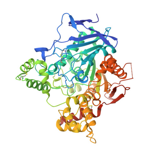
1ZGC

Crystal Structure of Torpedo Californica Acetylcholinesterase in Complex With an (RS)-Tacrine(10)-Hupyridone Inhibitor.


Experimental Data Snapshot

  • Method: X-RAY DIFFRACTION
  • Resolution: 2.10 Å
  • R-Value Free: 0.247 
  • R-Value Work: 0.196 
  • R-Value Observed: 0.199 

wwPDB Validation   3D Report Full Report


Ligand Structure Quality Assessment 


This is version 1.4 of the entry. See complete history


Literature

Crystal Packing Mediates Enantioselective Ligand Recognition at the Peripheral Site of Acetylcholinesterase

Haviv, H.Wong, D.M.Greenblatt, H.M.Carlier, P.R.Pang, Y.P.Silman, I.Sussman, J.L.

(2005) J Am Chem Soc 127: 11029-11036

  • DOI: https://doi.org/10.1021/ja051765f
  • Primary Citation of Related Structures:  
    1ZGB, 1ZGC

  • PubMed Abstract: 

    Recently, alkylene-linked heterodimers of tacrine (1) and 5-amino-5,6,7,8-tetrahydroquinolinone (2, hupyridone) were shown to exhibit higher acetylcholinesterase (AChE) inhibition than either monomeric 1 or 2. Such inhibitors are potential drug candidates for ameliorating the cognitive decrements in early Alzheimer patients. In an attempt to understand the inhibition mechanism of one such dimer, (RS)-(+/-)-N-9-(1,2,3,4-tetrahydroacridinyl)-N'-5-[5,6,7,8-tetrahydro-2'(1'H)-quinolinonyl]-1,10-diaminodecane [(RS)-(+/-)-3] bisoxalate, the racemate was soaked in trigonal Torpedo californica AChE (TcAChE) crystals, and the X-ray structure of the resulting complex was solved to 2.30 A resolution. Its structure revealed the 1 unit bound to the "anionic" subsite of the active site, near the bottom of the active-site gorge, as seen for the 1/TcAChE complex. Interestingly, only the (R)-enantiomer of the 2 unit was seen in the peripheral "anionic" site (PAS) at the top of the gorge, and was hydrogen-bonded to the side chains of residues belonging to an adjacent, symmetry-related AChE molecule covering the gorge entrance. When the same racemate was soaked in orthorhombic crystals of TcAChE, in which the entrance to the gorge is more exposed, the crystal structure of the corresponding complex revealed no substantial enantiomeric selectivity. This observation suggests that the apparent enantiomeric selectivity of trigonal crystals of TcAChE for (R)-3 is mainly due to crystal packing, resulting in preferential binding of one enantiomeric inhibitor both to its "host" enzyme and to its neighbor in the asymmetric unit, rather than to steric constraints imposed by the geometry of the active-site gorge.


  • Organizational Affiliation

    Departments of Structural Biology and Neurobiology, Weizmann Institute of Science, Rehovot 76100, Israel.


Macromolecules
Find similar proteins by:  (by identity cutoff)  |  3D Structure
Entity ID: 1
MoleculeChains Sequence LengthOrganismDetailsImage
Acetylcholinesterase
A, B
543Tetronarce californicaMutation(s): 0 
EC: 3.1.1.7
UniProt
Find proteins for P04058 (Tetronarce californica)
Explore P04058 
Go to UniProtKB:  P04058
Entity Groups  
Sequence Clusters30% Identity50% Identity70% Identity90% Identity95% Identity100% Identity
UniProt GroupP04058
Glycosylation
Glycosylation Sites: 3
Sequence Annotations
Expand
  • Reference Sequence
Small Molecules
Ligands 2 Unique
IDChains Name / Formula / InChI Key2D Diagram3D Interactions
A2E
Query on A2E

Download Ideal Coordinates CCD File 
F [auth A],
I [auth B]
(5S)-5-{[10-(1,2,3,4-TETRAHYDROACRIDIN-9-YLAMINO)DECYL]AMINO}-5,6,7,8-TETRAHYDROQUINOLIN-2(1H)-ONE
C32 H44 N4 O
ROTFGKJJMRTWBD-MHZLTWQESA-N
NAG
Query on NAG

Download Ideal Coordinates CCD File 
C [auth A],
D [auth A],
E [auth A],
G [auth B],
H [auth B]
2-acetamido-2-deoxy-beta-D-glucopyranose
C8 H15 N O6
OVRNDRQMDRJTHS-FMDGEEDCSA-N
Experimental Data & Validation

Experimental Data

  • Method: X-RAY DIFFRACTION
  • Resolution: 2.10 Å
  • R-Value Free: 0.247 
  • R-Value Work: 0.196 
  • R-Value Observed: 0.199 
  • Space Group: P 21 21 21
Unit Cell:
Length ( Å )Angle ( ˚ )
a = 90.996α = 90
b = 105.543β = 90
c = 150.448γ = 90
Software Package:
Software NamePurpose
REFMACrefinement
STRATEGYdata reduction
CCP4data scaling
XTALVIEWrefinement

Structure Validation

View Full Validation Report


Ligand Structure Quality Assessment 


Entry History 

Revision History  (Full details and data files)

  • Version 1.0: 2005-08-16
    Type: Initial release
  • Version 1.1: 2008-04-30
    Changes: Version format compliance
  • Version 1.2: 2011-07-13
    Changes: Non-polymer description, Version format compliance
  • Version 1.3: 2020-07-29
    Type: Remediation
    Reason: Carbohydrate remediation
    Changes: Data collection, Derived calculations, Structure summary
  • Version 1.4: 2024-10-30
    Changes: Data collection, Database references, Structure summary