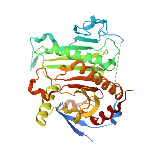
1Z84

X-ray structure of galt-like protein from arabidopsis thaliana at5g18200


Experimental Data Snapshot

  • Method: X-RAY DIFFRACTION
  • Resolution: 1.83 Å
  • R-Value Free: 0.231 
  • R-Value Work: 0.186 
  • R-Value Observed: 0.188 

Starting Model: experimental
View more details

wwPDB Validation   3D Report Full Report


Ligand Structure Quality Assessment 


This is version 1.5 of the entry. See complete history

Re-refinement Note

A newer entry is available that reflects an alternative modeling of the original data: 2Q4H


Literature

Structure and Mechanism of an ADP-Glucose Phosphorylase from Arabidopsis thaliana

MCCOY, J.G.ARABSHAHI, A.BITTO, E.BINGMAN, C.A.RUZICKA, F.J.FREY, P.A.PHILLIPS JR., G.N.

(2006) Biochemistry 45: 3154-3162

  • DOI: https://doi.org/10.1021/bi052232m
  • Primary Citation of Related Structures:  
    1Z84, 1ZWJ

  • PubMed Abstract: 

    The X-ray crystal structure of the At5g18200.1 protein has been determined to a nominal resolution of 2.30 A. The structure has a histidine triad (HIT)-like fold containing two distinct HIT-like motifs. The sequence of At5g18200.1 indicates a distant family relationship to the Escherichia coli galactose-1-P uridylyltransferase (GalT): the determined structure of the At5g18200.1 protein confirms this relationship. The At5g18200.1 protein does not demonstrate GalT activity but instead catalyzes adenylyl transfer in the reaction of ADP-glucose with various phosphates. The best acceptor among those evaluated is phosphate itself; thus, the At5g18200.1 enzyme appears to be an ADP-glucose phosphorylase. The enzyme catalyzes the exchange of (14)C between ADP-[(14)C]glucose and glucose-1-P in the absence of phosphate. The steady state kinetics of exchange follows the ping-pong bi-bi kinetic mechanism, with a k(cat) of 4.1 s(-)(1) and K(m) values of 1.4 and 83 microM for ADP-[(14)C]glucose and glucose-1-P, respectively, at pH 8.5 and 25 degrees C. The overall reaction of ADP-glucose with phosphate to produce ADP and glucose-1-P follows ping-pong bi-bi steady state kinetics, with a k(cat) of 2.7 s(-)(1) and K(m) values of 6.9 and 90 microM for ADP-glucose and phosphate, respectively, at pH 8.5 and 25 degrees C. The kinetics are consistent with a double-displacement mechanism that involves a covalent adenylyl-enzyme intermediate. The X-ray crystal structure of this intermediate was determined to 1.83 A resolution and shows the AMP group bonded to His(186). The value of K(eq) in the direction of ADP and glucose-1-P formation is 5.0 at pH 7.0 and 25 degrees C in the absence of a divalent metal ion, and it is 40 in the presence of 1 mM MgCl(2).


  • Organizational Affiliation

    Department of Biochemistry, University of Wisconsin, Madison, Wisconsin 53706, USA.


Macromolecules
Find similar proteins by:  (by identity cutoff)  |  3D Structure
Entity ID: 1
MoleculeChains Sequence LengthOrganismDetailsImage
galactose-1-phosphate uridyl transferase-like protein
A, B
351Arabidopsis thalianaMutation(s): 0 
Gene Names: AT5G18200
EC: 2.7.7
UniProt
Find proteins for Q9FK51 (Arabidopsis thaliana)
Explore Q9FK51 
Go to UniProtKB:  Q9FK51
Entity Groups  
Sequence Clusters30% Identity50% Identity70% Identity90% Identity95% Identity100% Identity
UniProt GroupQ9FK51
Sequence Annotations
Expand
  • Reference Sequence
Small Molecules
Ligands 3 Unique
IDChains Name / Formula / InChI Key2D Diagram3D Interactions
AMP
Query on AMP

Download Ideal Coordinates CCD File 
E [auth A],
K [auth B]
ADENOSINE MONOPHOSPHATE
C10 H14 N5 O7 P
UDMBCSSLTHHNCD-KQYNXXCUSA-N
ZN
Query on ZN

Download Ideal Coordinates CCD File 
C [auth A],
D [auth A],
I [auth B],
J [auth B]
ZINC ION
Zn
PTFCDOFLOPIGGS-UHFFFAOYSA-N
EDO
Query on EDO

Download Ideal Coordinates CCD File 
F [auth A]
G [auth A]
H [auth A]
L [auth B]
M [auth B]
F [auth A],
G [auth A],
H [auth A],
L [auth B],
M [auth B],
N [auth B]
1,2-ETHANEDIOL
C2 H6 O2
LYCAIKOWRPUZTN-UHFFFAOYSA-N
Experimental Data & Validation

Experimental Data

  • Method: X-RAY DIFFRACTION
  • Resolution: 1.83 Å
  • R-Value Free: 0.231 
  • R-Value Work: 0.186 
  • R-Value Observed: 0.188 
  • Space Group: P 21 21 21
Unit Cell:
Length ( Å )Angle ( ˚ )
a = 62.037α = 90
b = 95.65β = 90
c = 110.918γ = 90
Software Package:
Software NamePurpose
DENZOdata reduction
SCALEPACKdata scaling
MOLREPphasing
REFMACrefinement

Structure Validation

View Full Validation Report


Ligand Structure Quality Assessment 


Entry History 

Deposition Data

Revision History  (Full details and data files)

  • Version 1.0: 2005-04-19
    Type: Initial release
  • Version 1.1: 2008-04-30
    Changes: Version format compliance
  • Version 1.2: 2011-07-13
    Changes: Version format compliance
  • Version 1.3: 2017-10-11
    Changes: Refinement description
  • Version 1.4: 2023-08-23
    Changes: Data collection, Database references, Derived calculations, Refinement description
  • Version 1.5: 2024-10-16
    Changes: Structure summary