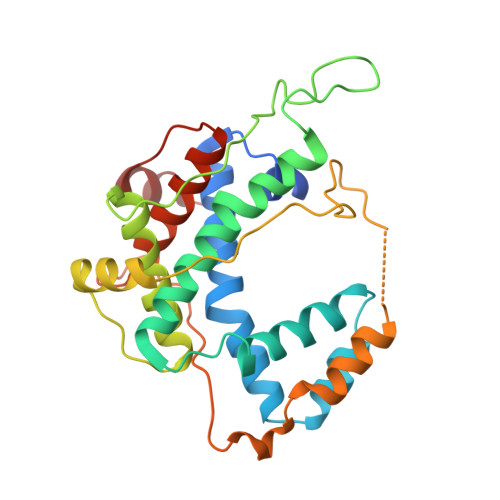
Crystal structure of a fragment of mouse ubiquitin-activating enzyme.
Szczepanowski, R.H., Filipek, R., Bochtler, M.(2005) J Biological Chem 280: 22006-22011
- PubMed: 15774460 Search on PubMed
- DOI: https://doi.org/10.1074/jbc.M502583200
- Primary Citation Related Structures:
1Z7L - PubMed Abstract:
Protein ubiquitination requires the sequential activity of three enzymes: a ubiquitin-activating enzyme (E1), a ubiquitin-conjugating enzyme (E2), and a ubiquitin-ligase (E3). The ubiquitin-transfer machinery is hierarchically organized; for every ubiquitin-activating enzyme, there are several ubiquitin-conjugating enzymes, and most ubiquitin-conjugating enzymes can in turn interact with multiple ubiquitin ligases. Despite the central role of ubiquitin-activating enzyme in this cascade, a crystal structure of a ubiquitin-activating enzyme is not available. The enzyme is thought to consist of an adenylation domain, a catalytic cysteine domain, a four-helix bundle, and possibly, a ubiquitin-like domain. Its adenylation domain can be modeled because it is clearly homologous to the structurally known adenylation domains of the activating enzymes for the small ubiquitin-like modifier (SUMO) and for the protein encoded by the neuronal precursor cell-expressed, developmentally down-regulated gene 8 (NEDD8). Low sequence similarity and vastly different domain lengths make modeling difficult for the catalytic cysteine domain that results from the juxtaposition of two catalytic cysteine half-domains. Here, we present a biochemical and crystallographic characterization of the two half-domains and the crystal structure of the larger, second catalytic cysteine half-domain of mouse ubiquitin-activating enzyme. We show that the domain is organized around a conserved folding motif that is also present in the NEDD8- and SUMO-activating enzymes, and we propose a tentative model for full-length ubiquitin-activating enzyme.
- International Institute of Molecular and Cell Biology, ul. Trojdena 4, 02-109 Warsaw, Poland.
Organizational Affiliation: