Encephalitozoon cuniculi mRNA cap (guanine N-7) methyltransferase: methyl acceptor specificity, inhibition BY S-adenosylmethionine analogs, and structure-guided mutational analysis.
Hausmann, S., Zheng, S., Fabrega, C., Schneller, S.W., Lima, C.D., Shuman, S.(2005) J Biol Chem 280: 20404-20412
- PubMed: 15760890 Search on PubMed
- DOI: https://doi.org/10.1074/jbc.M501073200
- Primary Citation Related Structures:
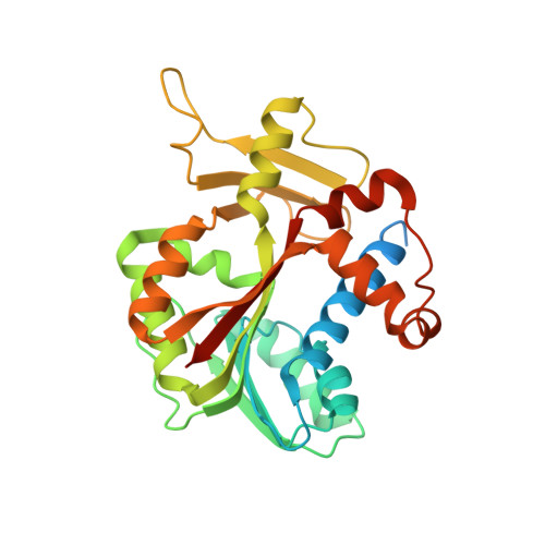
1Z3C - PubMed Abstract:
The Encephalitozoon cuniculi mRNA cap (guanine N-7) methyltransferase Ecm1 has been characterized structurally but not biochemically. Here we show that purified Ecm1 is a monomeric protein that catalyzes methyl transfer from S-adenosylmethionine (AdoMet) to GTP. The reaction is cofactor-independent and optimal at pH 7.5. Ecm1 also methylates GpppA, GDP, and dGTP but not ATP, CTP, UTP, ITP, or m(7)GTP. The affinity of Ecm1 for the cap dinucleotide GpppA (K 0.1 mm) is higher than that for GTP (K(m) 1 mm) or GDP (K(m) 2.4 mm). Methylation of GTP by Ecm1 in the presence of 5 microm AdoMet is inhibited by the reaction product AdoHcy (IC(50) 4 microm) and by substrate analogs sinefungin (IC(50) 1.5 microm), aza-AdoMet (IC(50) 100 microm), and carbocyclic aza-AdoMet (IC(50) 35 microm). The crystal structure of an Ecm1.aza-AdoMet binary complex reveals that the inhibitor occupies the same site as AdoMet. Structure-function analysis of Ecm1 by alanine scanning and conservative substitutions identified functional groups necessary for methyltransferase activity in vivo. Amino acids Lys-54, Asp-70, Asp-78, and Asp-94, which comprise the AdoMet-binding site, and Phe-141, which contacts the cap guanosine, are essential for cap methyltransferase activity in vitro.
- Molecular Biology and Structural Biology Programs, Sloan-Kettering Institute, New York, New York 10021, USA.
Organizational Affiliation: