1YYM | pdb_00001yym

crystal structure of F23, a scorpion-toxin mimic of CD4, in complex with HIV-1 YU2 gp120 envelope glycoprotein and anti-HIV-1 antibody 17b


Experimental Data Snapshot

  • Method: X-RAY DIFFRACTION
  • Resolution: 2.20 Å
  • R-Value Free: 
    0.268 (Depositor), 0.263 (DCC) 
  • R-Value Work: 
    0.209 (Depositor), 0.206 (DCC) 
  • R-Value Observed: 
    0.209 (Depositor) 

Starting Model: experimental
View more details

wwPDB Validation 3D Report Full Report

Validation slider image for 1YYM

Ligand Structure Quality Assessment 


This is version 2.1 of the entry. See complete history

Literature

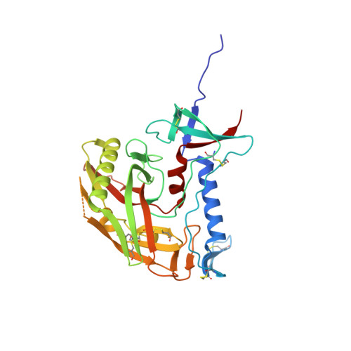
Scorpion-toxin mimics of CD4 in complex with human immunodeficiency virus gp120 crystal structures, molecular mimicry, and neutralization breadth.

Huang, C.C.Stricher, F.Martin, L.Decker, J.M.Majeed, S.Barthe, P.Hendrickson, W.A.Robinson, J.Roumestand, C.Sodroski, J.Wyatt, R.Shaw, G.M.Vita, C.Kwong, P.D.

(2005) Structure 13: 755-768

  • DOI: https://doi.org/10.1016/j.str.2005.03.006
  • Primary Citation Related Structures: 
    1YYL, 1YYM

  • PubMed Abstract: 

    The binding surface on CD4 for the HIV-1 gp120 envelope glycoprotein has been transplanted previously onto a scorpion-toxin scaffold. Here, we use X-ray crystallography to characterize atomic-level details of gp120 with this transplant, CD4M33. Despite known envelope flexibility, the conformation of gp120 induced by CD4M33 was so similar to that induced by CD4 that localized measures were required to distinguish ligand-induced differences from lattice variation. To investigate relationships between structure, function, and mimicry, an F23 analog of CD4M33 was devised. Structural and thermodynamic analyses showed F23 to be a better molecular mimic of CD4 than CD4M33. F23 also showed increased neutralization breadth, against diverse isolates of HIV-1, HIV-2, and SIVcpz. Our results lend insight into the stability of the CD4 bound conformation of gp120, define measures that quantify molecular mimicry as a function of evolutionary distance, and suggest how such evaluations might be useful in developing mimetic antagonists with increased neutralization breadth.


  • Organizational Affiliation
    • Vaccine Research Center, National Institutes of Health, Bethesda, Maryland 20892, USA.

Macromolecule Content 

  • Total Structure Weight: 174.9 kDa 
  • Atom Count: 12,823 
  • Modeled Residue Count: 1,540 
  • Deposited Residue Count: 1,566 
  • Unique protein chains: 4

Macromolecules

Find similar proteins by:|  3D Structure
Entity ID: 1
MoleculeChains  Sequence LengthOrganismDetailsImage
Exterior membrane glycoprotein(GP120),Exterior membrane glycoprotein(GP120),Exterior membrane glycoprotein(GP120)A [auth G],
E [auth P]
313Human immunodeficiency virus 1Mutation(s): 0 
UniProt
Find proteins for P35961 (Human immunodeficiency virus type 1 group M subtype B (isolate YU-2))
Explore P35961 
Go to UniProtKB:  P35961
Entity Groups
Sequence Clusters30% Identity50% Identity70% Identity90% Identity95% Identity100% Identity
UniProt GroupP35961
Glycosylation
Glycosylation Sites: 8
Sequence Annotations
Expand
Reference Sequence
Find similar proteins by:|  3D Structure
Entity ID: 2
MoleculeChains  Sequence LengthOrganismDetailsImage
antibody 17b light chainB [auth L],
F [auth Q]
214Homo sapiensMutation(s): 0 
Find similar proteins by:|  3D Structure
Entity ID: 3
MoleculeChains  Sequence LengthOrganismDetailsImage
antibody 17b heavy chainC [auth H],
G [auth R]
229Homo sapiensMutation(s): 0 
Find similar proteins by:|  3D Structure
Entity ID: 4
MoleculeChains  Sequence LengthOrganismDetailsImage
F23, scorpion-toxin mimic of CD4D [auth M],
H [auth S]
27synthetic constructMutation(s): 0 

Small Molecules

Ligands 3 Unique
IDChains Name / Formula / InChI Key2D Diagram3D Interactions
NAG

Query on NAG


Download:Ideal Coordinates CCD File
AA [auth P]
I [auth G]
J [auth G]
K [auth G]
L [auth G]
AA [auth P],
I [auth G],
J [auth G],
K [auth G],
L [auth G],
M [auth G],
N [auth G],
O [auth G],
P [auth G],
T [auth P],
U [auth P],
V [auth P],
W [auth P],
X [auth P],
Y [auth P],
Z [auth P]
2-acetamido-2-deoxy-beta-D-glucopyranose
C8 H15 N O6
OVRNDRQMDRJTHS-FMDGEEDCSA-N
EDO

Query on EDO


Download:Ideal Coordinates CCD File
Q [auth G],
S [auth M]
1,2-ETHANEDIOL
C2 H6 O2
LYCAIKOWRPUZTN-UHFFFAOYSA-N
IPA

Query on IPA


Download:Ideal Coordinates CCD File
BA [auth P],
R [auth G]
ISOPROPYL ALCOHOL
C3 H8 O
KFZMGEQAYNKOFK-UHFFFAOYSA-N

Experimental Data & Validation

Experimental Data

  • Method: X-RAY DIFFRACTION
  • Resolution: 2.20 Å
  • R-Value Free:  0.268 (Depositor), 0.263 (DCC) 
  • R-Value Work:  0.209 (Depositor), 0.206 (DCC) 
  • R-Value Observed: 0.209 (Depositor) 
Space Group: P 1 21 1
Unit Cell:
Length ( Å )Angle ( ˚ )
a = 51.62α = 90
b = 157.041β = 93.45
c = 108.929γ = 90
Software Package:
Software NamePurpose
CNSrefinement
HKL-2000data reduction
SCALEPACKdata scaling
CNSphasing

Structure Validation

View Full Validation Report


Ligand Structure Quality Assessment 


Entry History 

Revision History  (Full details and data files)

  • Version 1.0: 2005-05-03
    Type: Initial release
  • Version 1.1: 2008-04-30
    Changes: Version format compliance
  • Version 1.2: 2011-07-13
    Changes: Non-polymer description, Version format compliance
  • Version 1.3: 2017-06-14
    Changes: Database references, Source and taxonomy, Structure summary
  • Version 1.4: 2019-12-11
    Changes: Derived calculations
  • Version 2.0: 2020-07-29
    Type: Remediation
    Reason: Carbohydrate remediation
    Changes: Advisory, Atomic model, Data collection, Derived calculations, Non-polymer description, Structure summary
  • Version 2.1: 2023-10-25
    Changes: Data collection, Database references, Refinement description, Structure summary