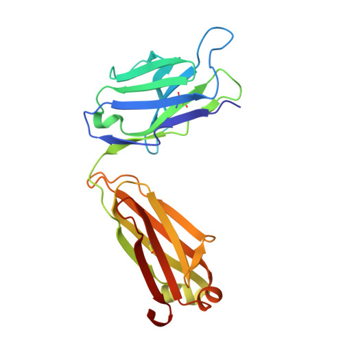
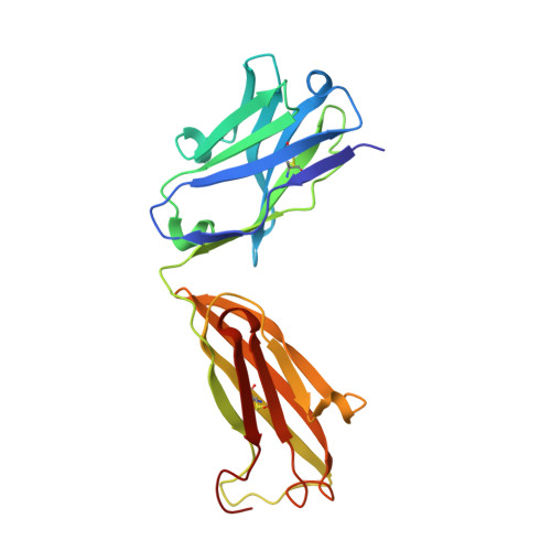
1YNL

Identification of Key residues of the NC6.8 Fab antibody fragment binding to synthetic sweeterners: Crystal structure of NC6.8 co-crystalized with high potency sweetener compound SC45647


Experimental Data Snapshot

  • Method: X-RAY DIFFRACTION
  • Resolution: 1.70 Å
  • R-Value Free: 0.241 
  • R-Value Work: 0.207 
  • R-Value Observed: 0.208 

wwPDB Validation   3D Report Full Report


Ligand Structure Quality Assessment 


This is version 1.5 of the entry. See complete history


Literature

Cocrystal Structures of NC6.8 Fab Identify Key Interactions for High Potency Sweetener Recognition: Implications for the Design of Synthetic Sweeteners

Gokulan, K.Khare, S.Ronning, D.R.Linthicum, S.D.Sacchettini, J.C.Rupp, B.

(2005) Biochemistry 44: 9889-9898

  • DOI: https://doi.org/10.1021/bi050613u
  • Primary Citation of Related Structures:  
    1YNK, 1YNL

  • PubMed Abstract: 

    The crystal structures of the murine monoclonal IgG2b(kappa) antibody NC6.8 Fab fragment complexed with high-potency sweetener compound SC45647 and nontasting high-affinity antagonist TES have been determined. The crystal structures show how sweetener potency is fine-tuned by multiple interactions between specific receptor residues and the functionally different groups of the sweeteners. Comparative analysis with the structure of NC6.8 complexed with the super-potency sweetener NC174 reveals that although the same residues in the antigen binding pocket of NC6.8 interact with the zwitterionic, trisubstituted guanidinium sweeteners as well as TES, specific differences exist and provide guidance for the design of new artificial sweeteners. In case of the nonsweetener TES, the interactions with the receptor are indirectly mediated through a hydrogen bonded water network, while the sweeteners bind with high affinity directly to the receptor. The presence of a hydrophobic group interacting with multiple receptor residues as a major determinant for sweet taste has been confirmed. The nature of the hydrophobic group is likely a discriminator for super- versus high-potency sweeteners, which can be exploited in the design of new, highly potent sweetener compounds. Overall similarities and partial conservation of interactions indicate that the NC6.8 Fab surrogate is representing crucial features of the T1R2 taste receptor VFTM binding site.


  • Organizational Affiliation

    Department of Biochemistry & Biophysics, Texas A&M University, College Station, Texas 77843-2128, USA.


Macromolecules
Find similar proteins by:  (by identity cutoff)  |  3D Structure
Entity ID: 1
MoleculeChains Sequence LengthOrganismDetailsImage
Ig gamma light chainA [auth L]219Mus musculusMutation(s): 0 
UniProt
Find proteins for A2P1G9 (Mus musculus)
Explore A2P1G9 
Go to UniProtKB:  A2P1G9
Entity Groups  
Sequence Clusters30% Identity50% Identity70% Identity90% Identity95% Identity100% Identity
UniProt GroupA2P1G9
Sequence Annotations
Expand
  • Reference Sequence
Find similar proteins by:  (by identity cutoff)  |  3D Structure
Entity ID: 2
MoleculeChains Sequence LengthOrganismDetailsImage
Ig gamma heavy chainB [auth H]219Mus musculusMutation(s): 0 
UniProt
Find proteins for Q6PJB2 (Mus musculus)
Explore Q6PJB2 
Go to UniProtKB:  Q6PJB2
Entity Groups  
Sequence Clusters30% Identity50% Identity70% Identity90% Identity95% Identity100% Identity
UniProt GroupQ6PJB2
Sequence Annotations
Expand
  • Reference Sequence
Small Molecules
Ligands 1 Unique
IDChains Name / Formula / InChI Key2D Diagram3D Interactions
NES
Query on NES

Download Ideal Coordinates CCD File 
C [auth L]2-(2-HYDROXY-1,1-DIHYDROXYMETHYL-ETHYLAMINO)-ETHANESULFONIC ACID
C6 H15 N O6 S
JOCBASBOOFNAJA-UHFFFAOYSA-N
Experimental Data & Validation

Experimental Data

  • Method: X-RAY DIFFRACTION
  • Resolution: 1.70 Å
  • R-Value Free: 0.241 
  • R-Value Work: 0.207 
  • R-Value Observed: 0.208 
  • Space Group: C 1 2 1
Unit Cell:
Length ( Å )Angle ( ˚ )
a = 136.226α = 90
b = 48.14β = 109.53
c = 76.09γ = 90
Software Package:
Software NamePurpose
REFMACrefinement
HKL-2000data reduction
SCALEPACKdata scaling
AMoREphasing

Structure Validation

View Full Validation Report


Ligand Structure Quality Assessment 


Entry History 

Deposition Data

Revision History  (Full details and data files)

  • Version 1.0: 2005-08-16
    Type: Initial release
  • Version 1.1: 2008-04-30
    Changes: Version format compliance
  • Version 1.2: 2011-07-13
    Changes: Version format compliance
  • Version 1.3: 2016-10-19
    Changes: Refinement description
  • Version 1.4: 2018-01-31
    Changes: Experimental preparation
  • Version 1.5: 2024-11-06
    Changes: Data collection, Database references, Derived calculations, Structure summary