1YKJ | pdb_00001ykj

A45G p-hydroxybenzoate hydroxylase with p-hydroxybenzoate bound


Experimental Data Snapshot

  • Method: X-RAY DIFFRACTION
  • Resolution: 2.00 Å
  • R-Value Free: 
    0.265 (Depositor), 0.266 (DCC) 
  • R-Value Work: 
    0.201 (Depositor), 0.202 (DCC) 
  • R-Value Observed: 
    0.201 (Depositor) 

Starting Model: experimental
View more details

wwPDB Validation 3D Report Full Report

Validation slider image for 1YKJ

Ligand Structure Quality Assessment 


This is version 1.5 of the entry. See complete history

Literature

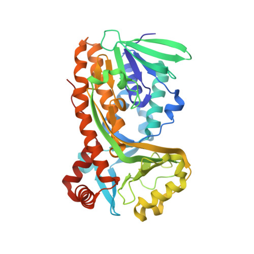
Removal of a methyl group causes global changes in p-hydroxybenzoate hydroxylase.

Cole, L.J.Gatti, D.L.Entsch, B.Ballou, D.P.

(2005) Biochemistry 44: 8047-8058

  • DOI: https://doi.org/10.1021/bi050108x
  • Primary Citation Related Structures: 
    1YKJ

  • PubMed Abstract: 

    p-Hydroxybenzoate hydroxylase (PHBH) is a homodimeric flavoprotein monooxygenase that catalyzes the hydroxylation of p-hydroxybenzoate to form 3,4-dihydroxybenzoate. Controlled catalysis is achieved by movement of the flavin and protein between three conformations, in, out, and open [Entsch, B., et al. (2005) Arch. Biochem. Biophys. 433, 297-311]. The open conformation is important for substrate binding and product release, the in conformation for reaction with oxygen and hydroxylation, and the out conformation for the reduction of FAD by NADPH. The open conformation is similar to the structure of Arg220Gln-PHBH in which the backbone peptide loop of residues 43-46, located on the si side of the flavin, is rotated. In this paper, we examine the structure and properties of the Ala45Gly-PHBH mutant enzyme. The crystal structure of the Ala45Gly enzyme is an asymmetric dimer, with one monomer similar (but not identical) to wild-type PHBH, while the other monomer has His72 flipped into solvent and replaced with Glu73 as one of several changes in the structure. The two structures correlate with evidence from kinetic studies for two forms of Ala45Gly-PHBH. One form of the enzyme dominates turnover and hydroxylates, while the other contributes little to turnover and fails to hydroxylate. Ala45Gly-PHBH favors the in conformation over alternative conformations. The effect of this mutation on the structure and function of PHBH illustrates the importance of the si side loop in the conformational state of PHBH and, consequently, the function of the enzyme. This work demonstrates some general principles of how enzymes use conformational movements to allow both access and egress of substrates and product, while restricting access to the solvent at a critical stage in catalysis.


  • Organizational Affiliation
    • Department of Biological Chemistry, University of Michigan, Ann Arbor, Michigan 48109-0606, USA.

Macromolecule Content 

  • Total Structure Weight: 92.09 kDa 
  • Atom Count: 6,958 
  • Modeled Residue Count: 786 
  • Deposited Residue Count: 788 
  • Unique protein chains: 1

Macromolecules

Find similar proteins by:|  3D Structure
Entity ID: 1
MoleculeChains  Sequence LengthOrganismDetailsImage
P-hydroxybenzoate hydroxylase
A, B
394Pseudomonas aeruginosaMutation(s): 1 
Gene Names: pobA
EC: 1.14.13.2
UniProt
Find proteins for P20586 (Pseudomonas aeruginosa (strain ATCC 15692 / DSM 22644 / CIP 104116 / JCM 14847 / LMG 12228 / 1C / PRS 101 / PAO1))
Explore P20586 
Go to UniProtKB:  P20586
Entity Groups
Sequence Clusters30% Identity50% Identity70% Identity90% Identity95% Identity100% Identity
UniProt GroupP20586
Sequence Annotations
Expand
Reference Sequence

Small Molecules

Ligands 4 Unique
IDChains Name / Formula / InChI Key2D Diagram3D Interactions
FAD

Query on FAD


Download:Ideal Coordinates CCD File
H [auth A],
Q [auth B]
FLAVIN-ADENINE DINUCLEOTIDE
C27 H33 N9 O15 P2
VWWQXMAJTJZDQX-UYBVJOGSSA-N
PSL

Query on PSL


Download:Ideal Coordinates CCD File
S [auth B],
T [auth B]
PYROSULFATE
O7 S2
VFNGKCDDZUSWLR-UHFFFAOYSA-L
PHB

Query on PHB


Download:Ideal Coordinates CCD File
I [auth A],
R [auth B]
P-HYDROXYBENZOIC ACID
C7 H6 O3
FJKROLUGYXJWQN-UHFFFAOYSA-N
SO4

Query on SO4


Download:Ideal Coordinates CCD File
C [auth A]
D [auth A]
E [auth A]
F [auth A]
G [auth A]
C [auth A],
D [auth A],
E [auth A],
F [auth A],
G [auth A],
J [auth B],
K [auth B],
L [auth B],
M [auth B],
N [auth B],
O [auth B],
P [auth B]
SULFATE ION
O4 S
QAOWNCQODCNURD-UHFFFAOYSA-L

Experimental Data & Validation

Experimental Data

  • Method: X-RAY DIFFRACTION
  • Resolution: 2.00 Å
  • R-Value Free:  0.265 (Depositor), 0.266 (DCC) 
  • R-Value Work:  0.201 (Depositor), 0.202 (DCC) 
  • R-Value Observed: 0.201 (Depositor) 
Space Group: P 21 21 2
Unit Cell:
Length ( Å )Angle ( ˚ )
a = 69.86α = 90
b = 85.897β = 90
c = 146.074γ = 90
Software Package:
Software NamePurpose
CNSrefinement
CrystalCleardata reduction
d*TREKdata scaling
CNSphasing

Structure Validation

View Full Validation Report


Ligand Structure Quality Assessment 


Entry History 

Deposition Data

Revision History  (Full details and data files)

  • Version 1.0: 2005-07-26
    Type: Initial release
  • Version 1.1: 2008-04-30
    Changes: Version format compliance
  • Version 1.2: 2011-07-13
    Changes: Version format compliance
  • Version 1.3: 2011-11-16
    Changes: Atomic model
  • Version 1.4: 2021-10-20
    Changes: Database references, Derived calculations
  • Version 1.5: 2023-08-23
    Changes: Data collection, Refinement description