1YHC | pdb_00001yhc

Crystal structure of Aquifex aeolicus LpxC deacetylase complexed with cacodylate


Experimental Data Snapshot

  • Method: X-RAY DIFFRACTION
  • Resolution: 2.10 Å
  • R-Value Free: 
    0.210 (Depositor) 
  • R-Value Work: 
    0.180 (Depositor) 
  • R-Value Observed: 
    0.182 (Depositor) 

Starting Model: experimental
View more details

wwPDB Validation 3D Report Full Report

Validation slider image for 1YHC

This is version 1.4 of the entry. See complete history

Literature

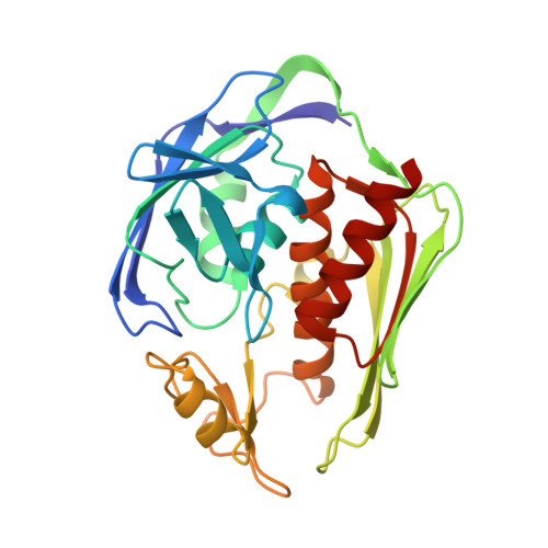
UDP-3-O-((R)-3-hydroxymyristoyl)-N-acetylglucosamine Deacetylase Functions through a General Acid-Base Catalyst Pair Mechanism

Hernick, M.Gennadios, H.A.Whittington, D.A.Rusche, K.M.Christianson, D.W.Fierke, C.A.

(2005) J Biological Chem 280: 16969-16978

  • DOI: https://doi.org/10.1074/jbc.M413560200
  • Primary Citation Related Structures: 
    1YH8, 1YHC

  • PubMed Abstract: 

    UDP-3-O-((R)-3-hydroxymyristoyl)-N-acetylglucosamine deacetylase (LpxC) is a zinc-dependent enzyme that catalyzes the deacetylation of UDP-3-O-((R)-3-hydroxymyristoyl)-N-acetylglucosamine to form UDP-3-O-(R-hydroxymyristoyl)glucosamine and acetate. The structural similarity of the active site of LpxC to metalloproteases led to the proposal that LpxC functions via a metalloprotease-like mechanism. The pH dependence of k(cat)/Km catalyzed by Escherichia coli and Aquifex aeolicus LpxC displayed a bell-shaped curve (EcLpxC yields apparent pKa values of 6.4+/-0.1 and 9.1+/-0.1), demonstrating that at least two ionizations are important for maximal activity. Metal substitution and mutagenesis experiments suggest that the basic limb of the pH profile is because of deprotonation of a zinc-coordinated group such as the zinc-water molecule, whereas the acidic limb of the pH profile is caused by protonation of either Glu78 or His265. Furthermore, the magnitude of the activity decreases and synergy observed for the active site mutants suggest that Glu78 and His265 act as a general acid-base catalyst pair. Crystal structures of LpxC complexed with cacodylate or palmitate demonstrate that both Glu78 and His265 hydrogen-bond with the same oxygen atom of the tetrahedral intermediate and the product carboxylate. These structural features suggest that LpxC catalyzes deacetylation by using Glu78 and His265 as a general acid-base pair and the zinc-bound water as a nucleophile.


  • Organizational Affiliation
    • Department of Chemistry, University of Michigan, Ann Arbor, Michigan 48109, USA.

Macromolecule Content 

  • Total Structure Weight: 63.22 kDa 
  • Atom Count: 4,721 
  • Modeled Residue Count: 532 
  • Deposited Residue Count: 540 
  • Unique protein chains: 1

Macromolecules

Find similar proteins by:|  3D Structure
Entity ID: 1
MoleculeChains  Sequence LengthOrganismDetailsImage
UDP-3-O-[3-hydroxymyristoyl] N-acetylglucosamine deacetylase
A, B
270Aquifex aeolicusMutation(s): 1 
Gene Names: lpxcenvA
EC: 3.5.1 (PDB Primary Data), 3.5.1.108 (UniProt)
UniProt
Find proteins for O67648 (Aquifex aeolicus (strain VF5))
Explore O67648 
Go to UniProtKB:  O67648
Entity Groups
Sequence Clusters30% Identity50% Identity70% Identity90% Identity95% Identity100% Identity
UniProt GroupO67648
Sequence Annotations
Expand
Reference Sequence

Small Molecules

Ligands 6 Unique
IDChains Name / Formula / InChI Key2D Diagram3D Interactions
PAM

Query on PAM


Download:Ideal Coordinates CCD File
J [auth A],
Q [auth B]
PALMITOLEIC ACID
C16 H30 O2
SECPZKHBENQXJG-FPLPWBNLSA-N
CAC

Query on CAC


Download:Ideal Coordinates CCD File
F [auth A],
N [auth B]
CACODYLATE ION
C2 H6 As O2
OGGXGZAMXPVRFZ-UHFFFAOYSA-M
SO4

Query on SO4


Download:Ideal Coordinates CCD File
G [auth A],
O [auth B]
SULFATE ION
O4 S
QAOWNCQODCNURD-UHFFFAOYSA-L
GOL

Query on GOL


Download:Ideal Coordinates CCD File
K [auth A]GLYCEROL
C3 H8 O3
PEDCQBHIVMGVHV-UHFFFAOYSA-N
ZN

Query on ZN


Download:Ideal Coordinates CCD File
C [auth A],
D [auth A],
E [auth A],
L [auth B],
M [auth B]
ZINC ION
Zn
PTFCDOFLOPIGGS-UHFFFAOYSA-N
CL

Query on CL


Download:Ideal Coordinates CCD File
H [auth A],
I [auth A],
P [auth B]
CHLORIDE ION
Cl
VEXZGXHMUGYJMC-UHFFFAOYSA-M

Experimental Data & Validation

Experimental Data

  • Method: X-RAY DIFFRACTION
  • Resolution: 2.10 Å
  • R-Value Free:  0.210 (Depositor) 
  • R-Value Work:  0.180 (Depositor) 
  • R-Value Observed: 0.182 (Depositor) 
Space Group: P 61
Unit Cell:
Length ( Å )Angle ( ˚ )
a = 101.279α = 90
b = 101.279β = 90
c = 122.745γ = 120
Software Package:
Software NamePurpose
HKL-2000data collection
SCALEPACKdata scaling
AMoREphasing
CNSrefinement
HKL-2000data reduction

Structure Validation

View Full Validation Report


Entry History 

Deposition Data

Revision History  (Full details and data files)

  • Version 1.0: 2005-02-15
    Type: Initial release
  • Version 1.1: 2008-04-30
    Changes: Version format compliance
  • Version 1.2: 2011-07-13
    Changes: Derived calculations, Version format compliance
  • Version 1.3: 2021-10-20
    Changes: Database references, Derived calculations
  • Version 1.4: 2023-08-23
    Changes: Data collection, Refinement description