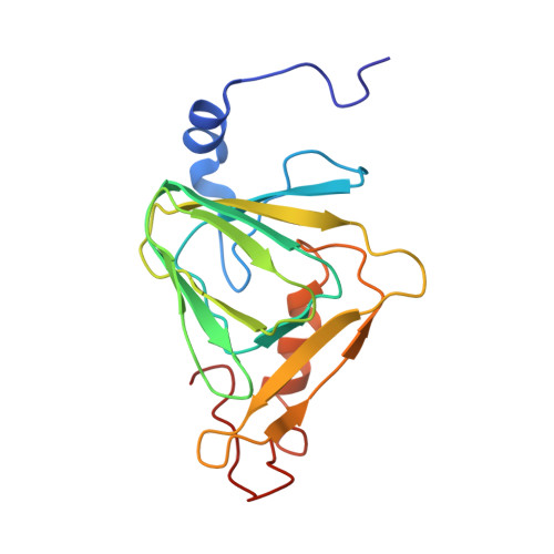
Structural Studies on 3-Hydroxyanthranilate-3,4-dioxygenase: The Catalytic Mechanism of a Complex Oxidation Involved in NAD Biosynthesis.
Zhang, Y., Colabroy, K.L., Begley, T.P., Ealick, S.E.(2005) Biochemistry 44: 7632-7643
- PubMed: 15909978
- DOI: https://doi.org/10.1021/bi047353l
- Primary Citation of Related Structures:
1YFU, 1YFW, 1YFX, 1YFY - PubMed Abstract:
3-Hydroxyanthranilate-3,4-dioxygenase (HAD) catalyzes the oxidative ring opening of 3-hydroxyanthranilate in the final enzymatic step of the biosynthetic pathway from tryptophan to quinolinate, the universal de novo precursor to the pyridine ring of nicotinamide adenine dinucleotide. The enzyme requires Fe2+ as a cofactor and is inactivated by 4-chloro-3-hydroxyanthranilate. HAD from Ralstonia metallidurans was crystallized, and the structure was determined at 1.9 A resolution. The structures of HAD complexed with the inhibitor 4-chloro-3-hydroxyanthranilic acid and either molecular oxygen or nitric oxide were determined at 2.0 A resolution, and the structure of HAD complexed with 3-hydroxyanthranilate was determined at 3.2 A resolution. HAD is a homodimer with a subunit topology that is characteristic of the cupin barrel fold. Each monomer contains two iron binding sites. The catalytic iron is buried deep inside the beta-barrel with His51, Glu57, and His95 serving as ligands. The other iron site forms an FeS4 center close to the solvent surface in which the sulfur atoms are provided by Cys125, Cys128, Cys162, and Cys165. The two iron sites are separated by 24 A. On the basis of the crystal structures of HAD, mutagenesis studies were carried out in order to elucidate the enzyme mechanism. In addition, a new mechanism for the enzyme inactivation by 4-chloro-3-hydroxyanthranilate is proposed.
- Department of Chemistry and Chemical Biology, Cornell University, Ithaca, New York 14853, USA.
Organizational Affiliation:


