1YBA | pdb_00001yba

The active form of phosphoglycerate dehydrogenase


Experimental Data Snapshot

  • Method: X-RAY DIFFRACTION
  • Resolution: 2.24 Å
  • R-Value Free: 
    0.235 (Depositor), 0.244 (DCC) 
  • R-Value Work: 
    0.186 (Depositor), 0.204 (DCC) 
  • R-Value Observed: 
    0.189 (Depositor) 

Starting Model: experimental
View more details

wwPDB Validation 3D Report Full Report

Validation slider image for 1YBA

Ligand Structure Quality Assessment 


This is version 1.4 of the entry. See complete history

Literature

Vmax Regulation through Domain and Subunit Changes. The Active Form of Phosphoglycerate Dehydrogenase

Thompson, J.R.Bell, J.K.Bratt, J.Grant, G.A.Banaszak, L.J.

(2005) Biochemistry 44: 5763-5773

  • DOI: https://doi.org/10.1021/bi047944b
  • Primary Citation Related Structures: 
    1YBA

  • PubMed Abstract: 

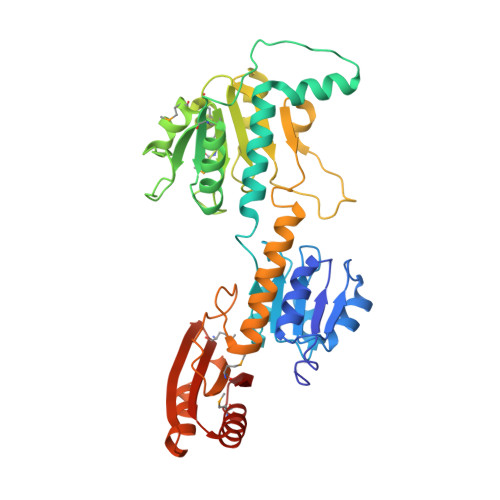
    An active conformation of phosphoglycerate dehydrogenase (PGDH) from Escherichia coli has been obtained using X-ray crystallography. The X-ray crystal structure is used to examine the potential intermediates for V(max) regulation, for the redox reaction, and for cooperative effects of serine binding. The crystal structure at 2.2 A resolution contains bound NAD(+) cofactor, either sulfate or phosphate anions, and alpha-ketoglutarate, a nonphysiological substrate. A PGDH subunit is formed from three distinct domains: regulatory (RBD), substrate (SBD), and nucleotide binding (NBD). The crystal conformation of the homotetramer points to the fact that, in the absence of serine, coordinated movement of the RBD-SBD domains occurs relative to the NBD. The result is a conformational change involving the steric relationships of both the domains and the subunits. Within the active site of each subunit is a bound molecule of alpha-ketoglutarate and the coenzyme, NAD. The catalytic or active site cleft is changed slightly although it is still solvent exposed; therefore, the catalytic reaction probably involves additional conformational changes. By comparing the inhibited with the uninhibited complex, it is possible to describe changes in conformation that are involved in the inhibitory signal transduction of serine.


  • Organizational Affiliation
    • Department of Biochemistry, Molecular Biology and Biophysics, University of Minnesota, Minneapolis, Minnesota 55455, USA.

Macromolecule Content 

  • Total Structure Weight: 182.12 kDa 
  • Atom Count: 14,017 
  • Modeled Residue Count: 1,624 
  • Deposited Residue Count: 1,640 
  • Unique protein chains: 1

Macromolecules

Find similar proteins by:|  3D Structure
Entity ID: 1
MoleculeChains  Sequence LengthOrganismDetailsImage
D-3-phosphoglycerate dehydrogenase
A, B, C, D
410Escherichia coliMutation(s): 8 
Gene Names: serA
EC: 1.1.1.95 (PDB Primary Data), 1.1.1.399 (UniProt)
UniProt
Find proteins for P0A9T0 (Escherichia coli (strain K12))
Explore P0A9T0 
Go to UniProtKB:  P0A9T0
Entity Groups
Sequence Clusters30% Identity50% Identity70% Identity90% Identity95% Identity100% Identity
UniProt GroupP0A9T0
Sequence Annotations
Expand
Reference Sequence

Small Molecules

Ligands 4 Unique
IDChains Name / Formula / InChI Key2D Diagram3D Interactions
NAD

Query on NAD


Download:Ideal Coordinates CCD File
J [auth A],
N [auth B],
S [auth C],
X [auth D]
NICOTINAMIDE-ADENINE-DINUCLEOTIDE
C21 H27 N7 O14 P2
BAWFJGJZGIEFAR-NNYOXOHSSA-N
AKG

Query on AKG


Download:Ideal Coordinates CCD File
G [auth A],
L [auth B],
P [auth C],
U [auth D]
2-OXOGLUTARIC ACID
C5 H6 O5
KPGXRSRHYNQIFN-UHFFFAOYSA-N
PO4

Query on PO4


Download:Ideal Coordinates CCD File
E [auth A],
F [auth A],
K [auth B],
O [auth C],
T [auth D]
PHOSPHATE ION
O4 P
NBIIXXVUZAFLBC-UHFFFAOYSA-K
UNL

Query on UNL


Download:Ideal Coordinates CCD File
H [auth A]
I [auth A]
M [auth B]
Q [auth C]
R [auth C]
H [auth A],
I [auth A],
M [auth B],
Q [auth C],
R [auth C],
V [auth D],
W [auth D]
Unknown ligand
NBIIXXVUZAFLBC-UHFFFAOYSA-K
Modified Residues  1 Unique
IDChains TypeFormula2D DiagramParent
MSE
Query on MSE
A, B, C, D
L-PEPTIDE LINKINGC5 H11 N O2 SeMET

Experimental Data & Validation

Experimental Data

  • Method: X-RAY DIFFRACTION
  • Resolution: 2.24 Å
  • R-Value Free:  0.235 (Depositor), 0.244 (DCC) 
  • R-Value Work:  0.186 (Depositor), 0.204 (DCC) 
  • R-Value Observed: 0.189 (Depositor) 
Space Group: P 43
Unit Cell:
Length ( Å )Angle ( ˚ )
a = 76.159α = 90
b = 76.159β = 90
c = 354.144γ = 90
Software Package:
Software NamePurpose
REFMACrefinement
DENZOdata reduction
SCALEPACKdata scaling
SOLVEphasing

Structure Validation

View Full Validation Report


Ligand Structure Quality Assessment 


Entry History 

Deposition Data

Revision History  (Full details and data files)

  • Version 1.0: 2005-04-26
    Type: Initial release
  • Version 1.1: 2008-04-02
    Changes: Version format compliance
  • Version 1.2: 2011-07-13
    Changes: Advisory, Refinement description, Version format compliance
  • Version 1.3: 2024-04-03
    Changes: Data collection, Database references, Derived calculations, Refinement description
  • Version 1.4: 2024-10-30
    Changes: Structure summary