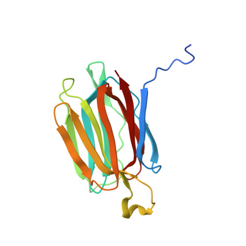
1XXR | pdb_00001xxr

Structure of a mannose-specific jacalin-related lectin from Morus Nigra in complex with mannose


Experimental Data Snapshot

  • Method: X-RAY DIFFRACTION
  • Resolution: 2.00 Å
  • R-Value Free: 
    0.211 (Depositor), 0.200 (DCC) 
  • R-Value Work: 
    0.191 (Depositor), 0.180 (DCC) 
  • R-Value Observed: 
    0.191 (Depositor) 

wwPDB Validation   3D Report Full Report


Ligand Structure Quality Assessment 

Created with Raphaël 2.3.0Worse 01 BetterLigand structure goodness of fit to experimental dataBest fitted MANClick on this verticalbar to view details

This is version 1.4 of the entry. See complete history


Literature

Structural analysis of the jacalin-related lectin MornigaM from the black mulberry (Morus nigra) in complex with mannose

Rabijns, A.Barre, A.Van Damme, E.J.M.Peumans, W.J.De Ranter, C.J.Rouge, P.

(2005) FEBS J 272: 3725-3732

  • DOI: https://doi.org/10.1111/j.1742-4658.2005.04801.x
  • Primary Citation of Related Structures:  
    1XXQ, 1XXR

  • PubMed Abstract: 

    The structures of MornigaM and the MornigaM-mannose complex have been determined at 1.8 A and 2.0 A resolution, respectively. Both structures adopt the typical beta-prism motif found in other jacalin-related lectins and their tetrameric assembly closely resembles that of jacalin. The carbohydrate-binding cavity of MornigaM readily binds mannose. No major structural rearrangements can be observed in MornigaM upon binding of mannose. These results allow corroboration of the structure-function relationships within the small group of Moraceae lectins.


  • Organizational Affiliation

    Laboratory of Analytical Chemistry and Medicinal Physicochemistry, Faculty of Pharmaceutical Sciences, K. U. Leuven, Belgium.


Macromolecules
Find similar proteins by:  (by identity cutoff)  |  3D Structure
Entity ID: 1
MoleculeChains Sequence LengthOrganismDetailsImage
mannose-binding lectin
A, B, C, D
161Morus nigraMutation(s): 0 
UniProt
Find proteins for Q8LGR3 (Morus nigra)
Explore Q8LGR3 
Go to UniProtKB:  Q8LGR3
Entity Groups  
Sequence Clusters30% Identity50% Identity70% Identity90% Identity95% Identity100% Identity
UniProt GroupQ8LGR3
Sequence Annotations
Expand
  • Reference Sequence
Small Molecules
Ligands 4 Unique
IDChains Name / Formula / InChI Key2D Diagram3D Interactions
MAN
Query on MAN

Download Ideal Coordinates CCD File 
G [auth B],
I [auth C],
K [auth D]
alpha-D-mannopyranose
C6 H12 O6
WQZGKKKJIJFFOK-PQMKYFCFSA-N
SO4
Query on SO4

Download Ideal Coordinates CCD File 
H [auth B]SULFATE ION
O4 S
QAOWNCQODCNURD-UHFFFAOYSA-L
GOL
Query on GOL

Download Ideal Coordinates CCD File 
F [auth A]GLYCEROL
C3 H8 O3
PEDCQBHIVMGVHV-UHFFFAOYSA-N
ACY
Query on ACY

Download Ideal Coordinates CCD File 
E [auth A],
J [auth C],
L [auth D]
ACETIC ACID
C2 H4 O2
QTBSBXVTEAMEQO-UHFFFAOYSA-N
Experimental Data & Validation

Experimental Data

  • Method: X-RAY DIFFRACTION
  • Resolution: 2.00 Å
  • R-Value Free:  0.211 (Depositor), 0.200 (DCC) 
  • R-Value Work:  0.191 (Depositor), 0.180 (DCC) 
  • R-Value Observed: 0.191 (Depositor) 
Space Group: P 65
Unit Cell:
Length ( Å )Angle ( ˚ )
a = 110.908α = 90
b = 110.908β = 90
c = 160.021γ = 120
Software Package:
Software NamePurpose
CNSrefinement
DENZOdata reduction
SCALEPACKdata scaling
X-PLORphasing

Structure Validation

View Full Validation Report


Ligand Structure Quality Assessment 

Created with Raphaël 2.3.0Worse 01 BetterLigand structure goodness of fit to experimental dataBest fitted MANClick on this verticalbar to view details

Entry History 

Deposition Data

Revision History  (Full details and data files)

  • Version 1.0: 2005-10-25
    Type: Initial release
  • Version 1.1: 2008-04-30
    Changes: Version format compliance
  • Version 1.2: 2011-07-13
    Changes: Non-polymer description, Version format compliance
  • Version 1.3: 2020-07-29
    Type: Remediation
    Reason: Carbohydrate remediation
    Changes: Data collection, Derived calculations, Structure summary
  • Version 1.4: 2024-03-13
    Changes: Data collection, Database references, Structure summary