1XWV | pdb_00001xwv

Structure of the house dust mite allergen Der f 2: Implications for function and molecular basis of IgE cross-reactivity


Experimental Data Snapshot

  • Method: X-RAY DIFFRACTION
  • Resolution: 1.83 Å
  • R-Value Free: 
    0.257 (Depositor), 0.237 (DCC) 
  • R-Value Work: 
    0.221 (Depositor), 0.228 (DCC) 
  • R-Value Observed: 
    0.221 (Depositor) 

Starting Model: experimental
View more details

wwPDB Validation 3D Report Full Report

Validation slider image for 1XWV

This is version 1.4 of the entry. See complete history

Literature

Structure of the house dust mite allergen Der f 2: implications for function and molecular basis of IgE cross-reactivity.

Johannessen, B.R.Skov, L.K.Kastrup, J.S.Kristensen, O.Bolwig, C.Larsen, J.N.Spangfort, M.Lund, K.Gajhede, M.

(2005) FEBS Lett 579: 1208-1212

  • DOI: https://doi.org/10.1016/j.febslet.2004.11.115
  • Primary Citation Related Structures: 
    1XWV

  • PubMed Abstract: 

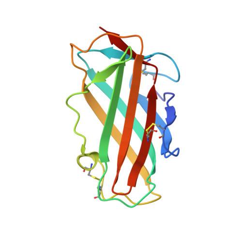
    The X-ray structure of the group 2 major allergen from Dermatophagoides farinae (Der f 2) was determined to 1.83 A resolution. The overall Der f 2 structure comprises a single domain of immunoglobulin fold with two anti-parallel beta-sheets. A large hydrophobic cavity is formed in the interior of Der f 2. Structural comparisons to distantly related proteins suggest a role in lipid binding. Immunoglobulin E (IgE) cross-reactivity between group 2 house dust mite major allergens can be explained by conserved surface areas representing IgE binding epitopes.


  • Organizational Affiliation
    • Biostructural Research, Department of Medicinal Chemistry, Danish University of Pharmaceutical Sciences, Universitetsparken 2, DK-2100 Copenhagen, Denmark.

Macromolecule Content 

  • Total Structure Weight: 29.2 kDa 
  • Atom Count: 2,329 
  • Modeled Residue Count: 258 
  • Deposited Residue Count: 258 
  • Unique protein chains: 1

Macromolecules

Find similar proteins by:|  3D Structure
Entity ID: 1
MoleculeChains  Sequence LengthOrganismDetailsImage
Der f II
A, B
129Dermatophagoides farinaeMutation(s): 0 
Membrane Entity: Yes 
UniProt
Find proteins for Q00855 (Dermatophagoides farinae)
Explore Q00855 
Go to UniProtKB:  Q00855
Entity Groups
Sequence Clusters30% Identity50% Identity70% Identity90% Identity95% Identity100% Identity
UniProt GroupQ00855
Sequence Annotations
Expand
Reference Sequence

Small Molecules

Ligands 2 Unique
IDChains Name / Formula / InChI Key2D Diagram3D Interactions
PE3

Query on PE3


Download:Ideal Coordinates CCD File
C [auth A]3,6,9,12,15,18,21,24,27,30,33,36,39-TRIDECAOXAHENTETRACONTANE-1,41-DIOL
C28 H58 O15
ILLKMACMBHTSHP-UHFFFAOYSA-N
XPE

Query on XPE


Download:Ideal Coordinates CCD File
D [auth B]3,6,9,12,15,18,21,24,27-NONAOXANONACOSANE-1,29-DIOL
C20 H42 O11
DTPCFIHYWYONMD-UHFFFAOYSA-N

Experimental Data & Validation

Experimental Data

  • Method: X-RAY DIFFRACTION
  • Resolution: 1.83 Å
  • R-Value Free:  0.257 (Depositor), 0.237 (DCC) 
  • R-Value Work:  0.221 (Depositor), 0.228 (DCC) 
  • R-Value Observed: 0.221 (Depositor) 
Space Group: P 1
Unit Cell:
Length ( Å )Angle ( ˚ )
a = 25.712α = 85.98
b = 47.638β = 75.9
c = 48.275γ = 82.76
Software Package:
Software NamePurpose
MOSFLMdata reduction
SCALAdata scaling
PHASERphasing
CNSrefinement
CCP4data scaling

Structure Validation

View Full Validation Report


Entry History 

Deposition Data

Revision History  (Full details and data files)

  • Version 1.0: 2004-12-14
    Type: Initial release
  • Version 1.1: 2008-04-30
    Changes: Version format compliance
  • Version 1.2: 2011-07-13
    Changes: Version format compliance
  • Version 1.3: 2023-10-25
    Changes: Data collection, Database references, Derived calculations, Refinement description
  • Version 1.4: 2024-11-06
    Changes: Structure summary