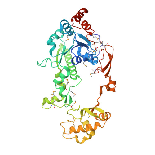
Crystal Structure of Escherichia coli Crotonobetainyl-CoA: Carnitine CoA-Transferase (CaiB) and Its Complexes with CoA and Carnitinyl-CoA.
Rangarajan, E.S., Li, Y., Iannuzzi, P., Cygler, M., Matte, A.(2005) Biochemistry 44: 5728-5738
- PubMed: 15823031 Search on PubMed
- DOI: https://doi.org/10.1021/bi047656f
- Primary Citation Related Structures:
1XK6, 1XK7, 1XVT, 1XVU, 1XVV - PubMed Abstract:
L-Carnitine (R-[-]-3-hydroxy-4-trimethylaminobutyrate) is found in both eukaryotic and prokaryotic cells and participates in diverse processes including long-chain fatty-acid transport and osmoprotection. The enzyme crotonobetainyl/gamma-butyrobetainyl-CoA:carnitine CoA-transferase (CaiB; E.C. 2.8.3.-) catalyzes the first step in carnitine metabolism, leading to the final product gamma-butyrobetaine. The crystal structures of Escherichia coli apo-CaiB, as well as its Asp169Ala mutant bound to CoA and to carnitinyl-CoA, have been determined and refined to 1.6, 2.4, and 2.4 A resolution, respectively. CaiB is composed of two identical circular chains that together form an intertwined dimer. Each monomer consists of a large domain, containing a Rossmann fold, and a small domain. The monomer and dimer resemble those of formyl-CoA transferase from Oxalobacter formigenes, as well as E. coli YfdW, a putative type-III CoA transferase of unknown function. The CoA cofactor-binding site is formed at the interface of the large domain of one monomer and the small domain from the second monomer. Most of the protein-CoA interactions are formed with the Rossmann fold domain. While the location of cofactor binding is similar in the three proteins, the specific CoA-protein interactions vary somewhat between CaiB, formyl-CoA transferase, and YfdW. CoA binding results in a change in the relative positions of the large and small domains compared with apo-CaiB. The observed carnitinyl-CoA product in crystals of the CaiB Asp169Ala mutant cocrystallized with crotonoyl-CoA and carnitine could result from (i) a catalytic mechanism involving a ternary enzyme-substrate complex, independent of a covalent anhydride intermediate with Asp169, (ii) a spontaneous reaction of the substrates in solution, followed by binding to the enzyme, or (iii) an involvement of another residue substituting functionally for Asp169, such as Glu23.
- Department of Biochemistry, McGill University, Montreal, Quebec, Canada.
Organizational Affiliation: