1XOW

Crystal structure of the human androgen receptor ligand binding domain bound with an androgen receptor NH2-terminal peptide, AR20-30, and R1881


Experimental Data Snapshot

  • Method: X-RAY DIFFRACTION
  • Resolution: 1.80 Å
  • R-Value Free: 0.213 
  • R-Value Work: 0.210 

wwPDB Validation   3D Report Full Report


Ligand Structure Quality Assessment 


This is version 1.5 of the entry. See complete history


Literature

Structural basis for androgen receptor interdomain and coactivator interactions suggests a transition in nuclear receptor activation function dominance

He, B.Gampe Jr., R.T.Kole, A.J.Hnat, A.T.Stanley, T.B.An, G.Stewart, E.L.Kalman, R.I.Minges, J.T.Wilson, E.M.

(2004) Mol Cell 16: 425-438

  • DOI: https://doi.org/10.1016/j.molcel.2004.09.036
  • Primary Citation of Related Structures:  
    1XOW, 1XQ3, 2AO6

  • PubMed Abstract: 

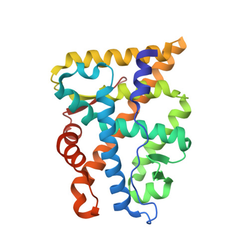
    The androgen receptor (AR) is required for male sex development and contributes to prostate cancer cell survival. In contrast to other nuclear receptors that bind the LXXLL motifs of coactivators, the AR ligand binding domain is preferentially engaged in an interdomain interaction with the AR FXXLF motif. Reported here are crystal structures of the ligand-activated AR ligand binding domain with and without bound FXXLF and LXXLL peptides. Key residues that establish motif binding specificity are identified through comparative structure-function and mutagenesis studies. A mechanism in prostate cancer is suggested by a functional AR mutation at a specificity-determining residue that recovers coactivator LXXLL motif binding. An activation function transition hypothesis is proposed in which an evolutionary decline in LXXLL motif binding parallels expansion and functional dominance of the NH(2)-terminal transactivation domain in the steroid receptor subfamily.


  • Organizational Affiliation

    Laboratories for Reproductive Biology, University of North Carolina at Chapel Hill, Chapel Hill, NC 27599, USA.


Macromolecules
Find similar proteins by:  (by identity cutoff)  |  3D Structure
Entity ID: 1
MoleculeChains Sequence LengthOrganismDetailsImage
androgen receptor249Homo sapiensMutation(s): 0 
Gene Names: ArNr3c4
UniProt & NIH Common Fund Data Resources
Find proteins for P10275 (Homo sapiens)
Explore P10275 
Go to UniProtKB:  P10275
PHAROS:  P10275
GTEx:  ENSG00000169083 
Entity Groups  
Sequence Clusters30% Identity50% Identity70% Identity90% Identity95% Identity100% Identity
UniProt GroupP10275
Sequence Annotations
Expand
  • Reference Sequence

Find similar proteins by:  Sequence   |   3D Structure  

Entity ID: 2
MoleculeChains Sequence LengthOrganismDetailsImage
decamer fragment of androgen receptor11N/AMutation(s): 0 
UniProt & NIH Common Fund Data Resources
Find proteins for P10275 (Homo sapiens)
Explore P10275 
Go to UniProtKB:  P10275
PHAROS:  P10275
GTEx:  ENSG00000169083 
Entity Groups  
Sequence Clusters30% Identity50% Identity70% Identity90% Identity95% Identity100% Identity
UniProt GroupP10275
Sequence Annotations
Expand
  • Reference Sequence
Small Molecules
Ligands 1 Unique
IDChains Name / Formula / InChI Key2D Diagram3D Interactions
R18
Query on R18

Download Ideal Coordinates CCD File 
C [auth A](17BETA)-17-HYDROXY-17-METHYLESTRA-4,9,11-TRIEN-3-ONE
C19 H24 O2
CCCIJQPRIXGQOE-XWSJACJDSA-N
Binding Affinity Annotations 
IDSourceBinding Affinity
R18 BindingDB:  1XOW Ki: 0.4 (nM) from 1 assay(s)
Kd: min: 0.52, max: 1.95 (nM) from 10 assay(s)
IC50: min: 0.3, max: 7 (nM) from 5 assay(s)
EC50: 0.01 (nM) from 1 assay(s)
PDBBind:  1XOW Kd: 0.52 (nM) from 1 assay(s)
Experimental Data & Validation

Experimental Data

  • Method: X-RAY DIFFRACTION
  • Resolution: 1.80 Å
  • R-Value Free: 0.213 
  • R-Value Work: 0.210 
  • Space Group: P 21 21 21
Unit Cell:
Length ( Å )Angle ( ˚ )
a = 54.941α = 90
b = 66.118β = 90
c = 70.44γ = 90
Software Package:
Software NamePurpose
MAR345data collection
HKL-2000data reduction
CNXrefinement
HKL-2000data scaling
CNXphasing

Structure Validation

View Full Validation Report


Ligand Structure Quality Assessment 


Entry History 

Deposition Data

Revision History  (Full details and data files)

  • Version 1.0: 2004-11-16
    Type: Initial release
  • Version 1.1: 2008-04-30
    Changes: Version format compliance
  • Version 1.2: 2011-07-13
    Changes: Version format compliance
  • Version 1.3: 2017-10-11
    Changes: Refinement description
  • Version 1.4: 2018-04-04
    Changes: Data collection
  • Version 1.5: 2023-08-23
    Changes: Data collection, Database references, Derived calculations, Refinement description