1XDQ | pdb_00001xdq

Structural and Biochemical Identification of a Novel Bacterial Oxidoreductase


Experimental Data Snapshot

  • Method: X-RAY DIFFRACTION
  • Resolution: 2.55 Å
  • R-Value Free: 
    0.274 (Depositor), 0.266 (DCC) 
  • R-Value Work: 
    0.230 (Depositor), 0.223 (DCC) 
  • R-Value Observed: 
    0.230 (Depositor) 

wwPDB Validation 3D Report Full Report

Validation slider image for 1XDQ

Ligand Structure Quality Assessment 


This is version 1.3 of the entry. See complete history

Literature

Structural and biochemical identification of a novel bacterial oxidoreductase.

Loschi, L.Brokx, S.J.Hills, T.L.Zhang, G.Bertero, M.G.Lovering, A.L.Weiner, J.H.Strynadka, N.C.

(2004) J Biological Chem 279: 50391-50400

  • DOI: https://doi.org/10.1074/jbc.M408876200
  • Primary Citation Related Structures: 
    1XDQ, 1XDY

  • PubMed Abstract: 

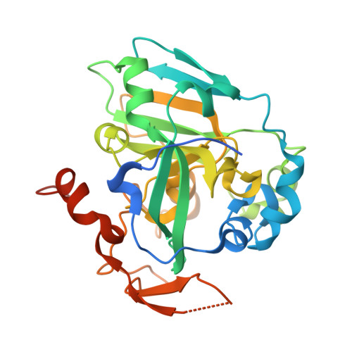
    By using a bioinformatics screen of the Escherichia coli genome for potential molybdenum-containing enzymes, we have identified a novel oxidoreductase conserved in the majority of Gram-negative bacteria. The identified operon encodes for a proposed heterodimer, YedYZ in Escherichia coli, consisting of a soluble catalytic subunit termed YedY, which is likely anchored to the membrane by a heme-containing trans-membrane subunit termed YedZ. YedY is uniquely characterized by the presence of one molybdenum molybdopterin not conjugated by an additional nucleotide, and it represents the only molybdoenzyme isolated from E. coli characterized by the presence of this cofactor form. We have further characterized the catalytic subunit YedY in both the molybdenum- and tungsten-substituted forms by using crystallographic analysis. YedY is very distinct in overall architecture from all known bacterial reductases but does show some similarity with the catalytic domain of the eukaryotic chicken liver sulfite oxidase. However, the strictly conserved residues involved in the metal coordination sphere and in the substrate binding pocket of YedY are strikingly different from that of chicken liver sulfite oxidase, suggesting a catalytic activity more in keeping with a reductase than that of a sulfite oxidase. Preliminary kinetic analysis of YedY with a variety of substrates supports our proposal that YedY and its many orthologues may represent a new type of membrane-associated bacterial reductase.


  • Organizational Affiliation
    • Department of Biochemistry, University of British Columbia, Vancouver, British Columbia V6T 1Z3, Canada.

Macromolecule Content 

  • Total Structure Weight: 171.17 kDa 
  • Atom Count: 10,771 
  • Modeled Residue Count: 1,313 
  • Deposited Residue Count: 1,490 
  • Unique protein chains: 1

Macromolecules

Find similar proteins by:|  3D Structure
Entity ID: 1
MoleculeChains  Sequence LengthOrganismDetailsImage
Bacterial Sulfite Oxidase
A, B, C, D, E
298Escherichia coliMutation(s): 0 
EC: 1.8.5
UniProt
Find proteins for P76342 (Escherichia coli (strain K12))
Explore P76342 
Go to UniProtKB:  P76342
Entity Groups
Sequence Clusters30% Identity50% Identity70% Identity90% Identity95% Identity100% Identity
UniProt GroupP76342
Sequence Annotations
Expand
Reference Sequence

Small Molecules

Ligands 4 Unique
IDChains Name / Formula / InChI Key2D Diagram3D Interactions
MTE

Query on MTE


Download:Ideal Coordinates CCD File
I [auth A],
M [auth B],
Q [auth C],
U [auth D],
Y [auth E]
PHOSPHONIC ACIDMONO-(2-AMINO-5,6-DIMERCAPTO-4-OXO-3,7,8A,9,10,10A-HEXAHYDRO-4H-8-OXA-1,3,9,10-TETRAAZA-ANTHRACEN-7-YLMETHYL)ESTER
C10 H14 N5 O6 P S2
HPEUEJRPDGMIMY-IFQPEPLCSA-N
MO

Query on MO


Download:Ideal Coordinates CCD File
G [auth A],
K [auth B],
O [auth C],
S [auth D],
W [auth E]
MOLYBDENUM ATOM
Mo
ZOKXTWBITQBERF-UHFFFAOYSA-N
URE

Query on URE


Download:Ideal Coordinates CCD File
F [auth A],
J [auth B],
N [auth C],
R [auth D],
V [auth E]
UREA
C H4 N2 O
XSQUKJJJFZCRTK-UHFFFAOYSA-N
O

Query on O


Download:Ideal Coordinates CCD File
H [auth A],
L [auth B],
P [auth C],
T [auth D],
X [auth E]
OXYGEN ATOM
O
XLYOFNOQVPJJNP-UHFFFAOYSA-N

Experimental Data & Validation

Experimental Data

  • Method: X-RAY DIFFRACTION
  • Resolution: 2.55 Å
  • R-Value Free:  0.274 (Depositor), 0.266 (DCC) 
  • R-Value Work:  0.230 (Depositor), 0.223 (DCC) 
  • R-Value Observed: 0.230 (Depositor) 
Space Group: I 21 21 21
Unit Cell:
Length ( Å )Angle ( ˚ )
a = 110.345α = 90
b = 165.064β = 90
c = 181.447γ = 90
Software Package:
Software NamePurpose
CNSrefinement
DENZOdata reduction
SCALEPACKdata scaling
SOLVEphasing

Structure Validation

View Full Validation Report


Ligand Structure Quality Assessment 


Entry History 

Deposition Data

Revision History  (Full details and data files)

  • Version 1.0: 2004-10-12
    Type: Initial release
  • Version 1.1: 2008-04-30
    Changes: Version format compliance
  • Version 1.2: 2011-07-13
    Changes: Version format compliance
  • Version 1.3: 2024-02-14
    Changes: Data collection, Database references, Derived calculations