1XCK | pdb_00001xck

Crystal structure of apo GroEL


Experimental Data Snapshot

  • Method: X-RAY DIFFRACTION
  • Resolution: 2.92 Å
  • R-Value Free: 
    0.235 (Depositor), 0.232 (DCC) 
  • R-Value Work: 
    0.203 (Depositor), 0.202 (DCC) 
  • R-Value Observed: 
    0.203 (Depositor) 

Starting Model: experimental
View more details

wwPDB Validation 3D Report Full Report

Validation slider image for 1XCK

This is version 1.3 of the entry. See complete history

Literature

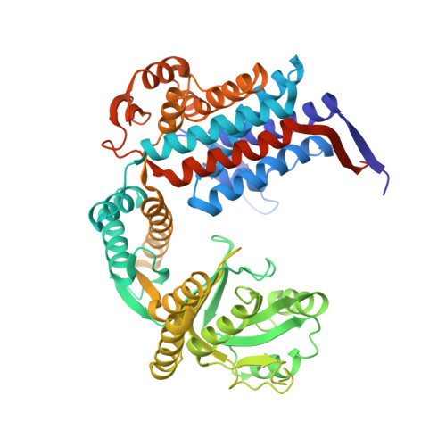
Crystal structure of wild-type chaperonin GroEL

Bartolucci, C.Lamba, D.Grazulis, S.Manakova, E.Heumann, H.

(2005) J Mol Biology 354: 940-951

  • DOI: https://doi.org/10.1016/j.jmb.2005.09.096
  • Primary Citation Related Structures: 
    1XCK

  • PubMed Abstract: 

    The 2.9A resolution crystal structure of apo wild-type GroEL was determined for the first time and represents the reference structure, facilitating the study of structural and functional differences observed in GroEL variants. Until now the crystal structure of the mutant Arg13Gly, Ala126Val GroEL was used for this purpose. We show that, due to the mutations as well as to the presence of a crystallographic symmetry, the ring-ring interface was inaccurately described. Analysis of the present structure allowed the definition of structural elements at this interface, essential for understanding the inter-ring allosteric signal transmission. We also show unambiguously that there is no ATP-induced 102 degrees rotation of the apical domain helix I around its helical axis, as previously assumed in the crystal structure of the (GroEL-KMgATP)(14) complex, and analyze the apical domain movements. These results enabled us to compare our structure with other GroEL crystal structures already published, allowing us to suggest a new route through which the allosteric signal for negative cooperativity propagates within the molecule. The proposed mechanism, supported by known mutagenesis data, underlines the importance of the switching of salt bridges.


  • Organizational Affiliation
    • Istituto di Cristallografia, CNR, P.O. Box 10, I-00016 Monterotondo Stazione Roma, Italy. cecilia.bartolucci@ic.cnr.it

Macromolecule Content 

  • Total Structure Weight: 810.09 kDa 
  • Atom Count: 55,301 
  • Modeled Residue Count: 7,336 
  • Deposited Residue Count: 7,658 
  • Unique protein chains: 1

Macromolecules

Find similar proteins by:|  3D Structure
Entity ID: 1
MoleculeChains  Sequence LengthOrganismDetailsImage
60 kDa chaperonin
A, B, C, D, E
A, B, C, D, E, F, G, H, I, J, K, L, M, N
547Escherichia coliMutation(s): 0 
EC: 5.6.1.7
UniProt
Find proteins for P0A6F5 (Escherichia coli (strain K12))
Explore P0A6F5 
Go to UniProtKB:  P0A6F5
Entity Groups
Sequence Clusters30% Identity50% Identity70% Identity90% Identity95% Identity100% Identity
UniProt GroupP0A6F5
Sequence Annotations
Expand
Reference Sequence

Small Molecules

Ligands 4 Unique
IDChains Name / Formula / InChI Key2D Diagram3D Interactions
MPD

Query on MPD


Download:Ideal Coordinates CCD File
BA [auth B]
CA [auth B]
DA [auth B]
EA [auth B]
EB [auth F]
BA [auth B],
CA [auth B],
DA [auth B],
EA [auth B],
EB [auth F],
EC [auth J],
FB [auth F],
GB [auth F],
IC [auth K],
JC [auth K],
KA [auth C],
LB [auth G],
MB [auth G],
NC [auth L],
RA [auth D],
RB [auth H],
SB [auth H],
SC [auth M],
T [auth A],
U [auth A],
VA [auth E],
WA [auth E],
WB [auth I],
WC [auth N],
XA [auth E],
XB [auth I],
XC [auth N],
YA [auth E],
YB [auth I],
YC [auth N],
ZA [auth E]
(4S)-2-METHYL-2,4-PENTANEDIOL
C6 H14 O2
SVTBMSDMJJWYQN-YFKPBYRVSA-N
PEG

Query on PEG


Download:Ideal Coordinates CCD File
AB [auth E]DI(HYDROXYETHYL)ETHER
C4 H10 O3
MTHSVFCYNBDYFN-UHFFFAOYSA-N
SO4

Query on SO4


Download:Ideal Coordinates CCD File
AC [auth J]
BB [auth F]
BC [auth J]
CB [auth F]
CC [auth J]
AC [auth J],
BB [auth F],
BC [auth J],
CB [auth F],
CC [auth J],
FA [auth C],
FC [auth K],
GA [auth C],
GC [auth K],
HA [auth C],
HB [auth G],
IA [auth C],
IB [auth G],
JB [auth G],
KC [auth L],
LA [auth D],
LC [auth L],
MA [auth D],
NA [auth D],
NB [auth H],
O [auth A],
OA [auth D],
OB [auth H],
OC [auth M],
P [auth A],
PA [auth D],
PB [auth H],
PC [auth M],
Q [auth A],
QC [auth M],
R [auth A],
SA [auth E],
TA [auth E],
TB [auth I],
TC [auth N],
UB [auth I],
UC [auth N],
V [auth B],
W [auth B],
X [auth B],
Y [auth B],
Z [auth B],
ZB [auth J]
SULFATE ION
O4 S
QAOWNCQODCNURD-UHFFFAOYSA-L
K

Query on K


Download:Ideal Coordinates CCD File
AA [auth B]
DB [auth F]
DC [auth J]
HC [auth K]
JA [auth C]
AA [auth B],
DB [auth F],
DC [auth J],
HC [auth K],
JA [auth C],
KB [auth G],
MC [auth L],
QA [auth D],
QB [auth H],
RC [auth M],
S [auth A],
UA [auth E],
VB [auth I],
VC [auth N]
POTASSIUM ION
K
NPYPAHLBTDXSSS-UHFFFAOYSA-N

Experimental Data & Validation

Experimental Data

  • Method: X-RAY DIFFRACTION
  • Resolution: 2.92 Å
  • R-Value Free:  0.235 (Depositor), 0.232 (DCC) 
  • R-Value Work:  0.203 (Depositor), 0.202 (DCC) 
  • R-Value Observed: 0.203 (Depositor) 
Space Group: P 21 21 21
Unit Cell:
Length ( Å )Angle ( ˚ )
a = 262.8α = 90
b = 283.6β = 90
c = 135.72γ = 90
Software Package:
Software NamePurpose
DENZOdata reduction
SCALEPACKdata scaling
AMoREphasing
CNSrefinement

Structure Validation

View Full Validation Report


Entry History 

Deposition Data

Revision History  (Full details and data files)

  • Version 1.0: 2005-10-25
    Type: Initial release
  • Version 1.1: 2008-04-30
    Changes: Version format compliance
  • Version 1.2: 2011-07-13
    Changes: Version format compliance
  • Version 1.3: 2023-10-25
    Changes: Data collection, Database references, Derived calculations, Refinement description