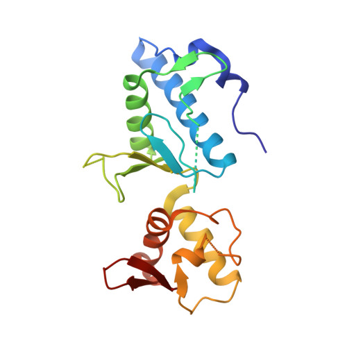
1XB4

Crystal structure of subunit VPS25 of the endosomal trafficking complex ESCRT-II


Experimental Data Snapshot

  • Method: X-RAY DIFFRACTION
  • Resolution: 3.10 Å
  • R-Value Free: 0.327 
  • R-Value Work: 0.327 
  • R-Value Observed: 0.273 

wwPDB Validation   3D Report Full Report


This is version 1.4 of the entry. See complete history


Literature

Crystal structure of subunit VPS25 of the endosomal trafficking complex ESCRT-II.

Wernimont, A.K.Weissenhorn, W.

(2004) BMC Struct Biol 4: 10-10

  • DOI: https://doi.org/10.1186/1472-6807-4-10
  • Primary Citation of Related Structures:  
    1XB4

  • PubMed Abstract: 

    Down-regulation of plasma membrane receptors via the endocytic pathway involves their monoubiquitylation, transport to endosomal membranes and eventual sorting into multi vesicular bodies (MVB) destined for lysosomal degradation. Successive assemblies of Endosomal Sorting Complexes Required for Transport (ESCRT-I, -II and III) largely mediate sorting of plasma membrane receptors at endosomal membranes, the formation of multivesicular bodies and their release into the endosomal lumen. In addition, the human ESCRT-II has been shown to form a complex with RNA polymerase II elongation factor ELL in order to exert transcriptional control activity. Here we report the crystal structure of Vps25 at 3.1 A resolution. Vps25 crystallizes in a dimeric form and each monomer is composed of two winged helix domains arranged in tandem. Structural comparisons detect no conformational changes between unliganded Vps25 and Vps25 within the ESCRT-II complex composed of two Vps25 copies and one copy each of Vps22 and Vps36 12. Our structural analyses present a framework for studying Vps25 interactions with ESCRT-I and ESCRT-III partners. Winged helix domain containing proteins have been implicated in nucleic acid binding and it remains to be determined whether Vps25 has a similar activity which might play a role in the proposed transcriptional control exerted by Vps25 and/or the whole ESCRT-II complex.


  • Organizational Affiliation

    European Molecular Biology Laboratory, Grenoble, France. wernimont@embl-grenoble.fr


Macromolecules
Find similar proteins by:  (by identity cutoff)  |  3D Structure
Entity ID: 1
MoleculeChains Sequence LengthOrganismDetailsImage
Hypothetical 23.6 kDa protein in YUH1-URA8 intergenic region
A, B, C, D
202Saccharomyces cerevisiaeMutation(s): 0 
Gene Names: YJR102CJ1957
UniProt
Find proteins for P47142 (Saccharomyces cerevisiae (strain ATCC 204508 / S288c))
Explore P47142 
Go to UniProtKB:  P47142
Entity Groups  
Sequence Clusters30% Identity50% Identity70% Identity90% Identity95% Identity100% Identity
UniProt GroupP47142
Sequence Annotations
Expand
  • Reference Sequence
Experimental Data & Validation

Experimental Data

  • Method: X-RAY DIFFRACTION
  • Resolution: 3.10 Å
  • R-Value Free: 0.327 
  • R-Value Work: 0.327 
  • R-Value Observed: 0.273 
  • Space Group: P 21 21 21
Unit Cell:
Length ( Å )Angle ( ˚ )
a = 53.36α = 90
b = 123.66β = 90
c = 140.3γ = 90
Software Package:
Software NamePurpose
ProDCdata collection
XDSdata reduction
SOLVEphasing
CNSrefinement
XDSdata scaling

Structure Validation

View Full Validation Report


Entry History 

Deposition Data

Revision History  (Full details and data files)

  • Version 1.0: 2005-03-22
    Type: Initial release
  • Version 1.1: 2008-04-30
    Changes: Version format compliance
  • Version 1.2: 2011-07-13
    Changes: Version format compliance
  • Version 1.3: 2017-10-11
    Changes: Refinement description
  • Version 1.4: 2024-02-14
    Changes: Data collection, Database references