1X6N | pdb_00001x6n

Crystal structure of S. marcescens chitinase A mutant W167A in complex with allosamidin


Experimental Data Snapshot

  • Method: X-RAY DIFFRACTION
  • Resolution: 2.00 Å
  • R-Value Free: 
    0.238 (Depositor) 
  • R-Value Work: 
    0.214 (Depositor), 0.230 (DCC) 
  • R-Value Observed: 
    0.214 (Depositor) 

Starting Model: experimental
View more details

wwPDB Validation 3D Report Full Report

Validation slider image for 1X6N

Ligand Structure Quality Assessment 


This is version 2.2 of the entry. See complete history

Literature

Muation of Trp167 at the -3 subsite of the chitin-binding cleft of S. marcescens chitinase A causes enhanced transglycosylation

Aronson Jr., N.N.Halloran, B.A.Alexyev, M.F.Zhou, X.E.Wang, Y.Meehan, E.J.Chen, L.

To be published.

Macromolecule Content 

  • Total Structure Weight: 61.61 kDa 
  • Atom Count: 4,347 
  • Modeled Residue Count: 539 
  • Deposited Residue Count: 563 
  • Unique protein chains: 1

Macromolecules

Find similar proteins by:|  3D Structure
Entity ID: 1
MoleculeChains  Sequence LengthOrganismDetailsImage
Chitinase A563Serratia marcescensMutation(s): 1 
Gene Names: chiA
EC: 3.2.1.14
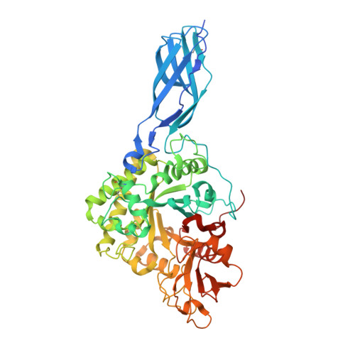
UniProt
Find proteins for P07254 (Serratia marcescens)
Explore P07254 
Go to UniProtKB:  P07254
Entity Groups
Sequence Clusters30% Identity50% Identity70% Identity90% Identity95% Identity100% Identity
UniProt GroupP07254
Sequence Annotations
Expand
Reference Sequence

Oligosaccharides

Help  
Entity ID: 2
MoleculeChains Length2D Diagram GlycosylationD Interactions
2-acetamido-2-deoxy-beta-D-allopyranose-(1-4)-2-acetamido-2-deoxy-beta-D-allopyranose
B
2N/A
Glycosylation Resources
GlyTouCan: G77950XB
GlyCosmos: G77950XB

Small Molecules

Ligands 1 Unique
IDChains Name / Formula / InChI Key2D Diagram3D Interactions
AMI

Query on AMI


Download:Ideal Coordinates CCD File
C [auth A]ALLOSAMIZOLINE
C9 H16 N2 O4
MKJAYSJDHSEFRI-PVFLNQBWSA-N

Biologically Interesting Molecules (External Reference) 

1 Unique

Experimental Data & Validation

Experimental Data

  • Method: X-RAY DIFFRACTION
  • Resolution: 2.00 Å
  • R-Value Free:  0.238 (Depositor) 
  • R-Value Work:  0.214 (Depositor), 0.230 (DCC) 
  • R-Value Observed: 0.214 (Depositor) 
Space Group: I 2 2 2
Unit Cell:
Length ( Å )Angle ( ˚ )
a = 76.67α = 90
b = 133.07β = 90
c = 192.22γ = 90
Software Package:
Software NamePurpose
CNSrefinement
HKL-2000data reduction
SCALEPACKdata scaling
CNSphasing

Structure Validation

View Full Validation Report


Ligand Structure Quality Assessment 


Entry History 

Deposition Data

Revision History  (Full details and data files)

  • Version 1.0: 2005-07-26
    Type: Initial release
  • Version 1.1: 2008-04-30
    Changes: Version format compliance
  • Version 1.2: 2011-07-13
    Changes: Version format compliance
  • Version 1.3: 2016-11-09
    Changes: Non-polymer description
  • Version 1.4: 2021-10-20
    Changes: Advisory, Database references, Structure summary
  • Version 2.0: 2022-04-13
    Changes: Atomic model, Data collection, Derived calculations, Non-polymer description, Structure summary
  • Version 2.1: 2023-08-23
    Changes: Data collection, Refinement description
  • Version 2.2: 2024-11-20
    Changes: Structure summary