1X5A | pdb_00001x5a

The solution structure of the second fibronectin type III domain of mouse Ephrin type-A receptor 1


Experimental Data Snapshot

  • Method: SOLUTION NMR
  • Conformers Calculated: 100 
  • Conformers Submitted: 20 
  • Selection Criteria: target function,structures with the least restraint violations 

wwPDB Validation 3D Report Full Report

Validation slider image for 1X5A

This is version 1.4 of the entry. See complete history

Literature

The solution structure of the second fibronectin type III domain of mouse Ephrin type-A receptor 1

Tochio, N.Sasagawa, A.Koshiba, S.Inoue, M.Kigawa, T.Yokoyama, S.

To be published.

Macromolecule Content 

  • Total Structure Weight: 11.67 kDa 
  • Atom Count: 822 
  • Modeled Residue Count: 107 
  • Deposited Residue Count: 107 
  • Unique protein chains: 1

Macromolecules

Find similar proteins by:|  3D Structure
Entity ID: 1
MoleculeChains  Sequence LengthOrganismDetailsImage
Ephrin type-A receptor 1107Mus musculusMutation(s): 0 
Gene Names: Epha1
EC: 2.7.1.112 (PDB Primary Data), 2.7.10.1 (UniProt)
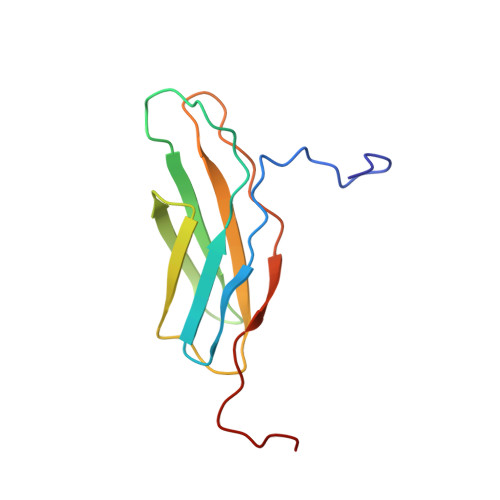
UniProt
Find proteins for Q60750 (Mus musculus)
Explore Q60750 
Go to UniProtKB:  Q60750
Entity Groups
Sequence Clusters30% Identity50% Identity70% Identity90% Identity95% Identity100% Identity
UniProt GroupQ60750
Sequence Annotations
Expand
Reference Sequence

Experimental Data & Validation

Experimental Data

  • Method: SOLUTION NMR
  • Conformers Calculated: 100 
  • Conformers Submitted: 20 
  • Selection Criteria: target function,structures with the least restraint violations 

Structure Validation

View Full Validation Report


Entry History 

Revision History  (Full details and data files)

  • Version 1.0: 2005-11-15
    Type: Initial release
  • Version 1.1: 2008-04-30
    Changes: Version format compliance
  • Version 1.2: 2011-07-13
    Changes: Version format compliance
  • Version 1.3: 2022-03-02
    Changes: Data collection, Database references, Derived calculations
  • Version 1.4: 2024-05-29
    Changes: Data collection