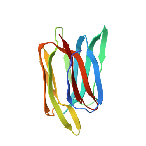
1X1V

Structure Of Banana Lectin- Methyl-Alpha-Mannose Complex


Experimental Data Snapshot

  • Method: X-RAY DIFFRACTION
  • Resolution: 2.45 Å
  • R-Value Free: 0.260 
  • R-Value Work: 0.224 
  • R-Value Observed: 0.226 

wwPDB Validation   3D Report Full Report


Ligand Structure Quality Assessment 


This is version 1.4 of the entry. See complete history


Literature

Unusual sugar specificity of banana lectin from Musa paradisiaca and its probable evolutionary origin. Crystallographic and modelling studies

Singh, D.D.Saikrishnan, K.Kumar, P.Surolia, A.Sekar, K.Vijayan, M.

(2005) Glycobiology 15: 1025-1032

  • DOI: https://doi.org/10.1093/glycob/cwi087
  • Primary Citation of Related Structures:  
    1X1V

  • PubMed Abstract: 

    The crystal structure of a complex of methyl-alpha-D-mannoside with banana lectin from Musa paradisiaca reveals two primary binding sites in the lectin, unlike in other lectins with beta-prism I fold which essentially consists of three Greek key motifs. It has been suggested that the fold evolved through successive gene duplication and fusion of an ancestral Greek key motif. In other lectins, all from dicots, the primary binding site exists on one of the three motifs in the three-fold symmetric molecule. Banana is a monocot, and the three motifs have not diverged enough to obliterate sequence similarity among them. Two Greek key motifs in it carry one primary binding site each. A common secondary binding site exists on the third Greek key. Modelling shows that both the primary sites can support 1-2, 1-3, and 1-6 linked mannosides with the second residue interacting in each case primarily with the secondary binding site. Modelling also readily leads to a bound branched mannopentose with the nonreducing ends of the two branches anchored at the two primary binding sites, providing a structural explanation for the lectin's specificity for branched alpha-mannans. A comparison of the dimeric banana lectin with other beta-prism I fold lectins, provides interesting insights into the variability in their quaternary structure.


  • Organizational Affiliation

    Molecular Biophysics Unit, Indian Institute of Science, Bangalore 560 012, Karnataka, India.


Macromolecules
Find similar proteins by:  (by identity cutoff)  |  3D Structure
Entity ID: 1
MoleculeChains Sequence LengthOrganismDetailsImage
lectin
A, B
141Musa acuminataMutation(s): 0 
UniProt
Find proteins for Q8L5H4 (Musa acuminata)
Explore Q8L5H4 
Go to UniProtKB:  Q8L5H4
Entity Groups  
Sequence Clusters30% Identity50% Identity70% Identity90% Identity95% Identity100% Identity
UniProt GroupQ8L5H4
Sequence Annotations
Expand
  • Reference Sequence
Small Molecules
Ligands 3 Unique
IDChains Name / Formula / InChI Key2D Diagram3D Interactions
MMA
Query on MMA

Download Ideal Coordinates CCD File 
C [auth A],
D [auth A],
E [auth A],
K [auth B],
L [auth B]
methyl alpha-D-mannopyranoside
C7 H14 O6
HOVAGTYPODGVJG-VEIUFWFVSA-N
HEZ
Query on HEZ

Download Ideal Coordinates CCD File 
H [auth A]
I [auth A]
J [auth A]
N [auth B]
O [auth B]
H [auth A],
I [auth A],
J [auth A],
N [auth B],
O [auth B],
P [auth B]
HEXANE-1,6-DIOL
C6 H14 O2
XXMIOPMDWAUFGU-UHFFFAOYSA-N
ZN
Query on ZN

Download Ideal Coordinates CCD File 
F [auth A],
G [auth A],
M [auth B]
ZINC ION
Zn
PTFCDOFLOPIGGS-UHFFFAOYSA-N
Experimental Data & Validation

Experimental Data

  • Method: X-RAY DIFFRACTION
  • Resolution: 2.45 Å
  • R-Value Free: 0.260 
  • R-Value Work: 0.224 
  • R-Value Observed: 0.226 
  • Space Group: P 32 2 1
Unit Cell:
Length ( Å )Angle ( ˚ )
a = 80.82α = 90
b = 80.82β = 90
c = 148γ = 120
Software Package:
Software NamePurpose
REFMACrefinement
DENZOdata reduction
SCALEPACKdata scaling
PHASERphasing

Structure Validation

View Full Validation Report


Ligand Structure Quality Assessment 


Entry History 

Deposition Data

Revision History  (Full details and data files)

  • Version 1.0: 2005-11-08
    Type: Initial release
  • Version 1.1: 2008-04-30
    Changes: Version format compliance
  • Version 1.2: 2011-07-13
    Changes: Advisory, Refinement description, Version format compliance
  • Version 1.3: 2020-07-29
    Type: Remediation
    Reason: Carbohydrate remediation
    Changes: Advisory, Data collection, Derived calculations, Structure summary
  • Version 1.4: 2023-10-25
    Changes: Data collection, Database references, Refinement description, Structure summary