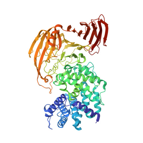
1X1J

Crystal Structure of Xanthan Lyase (N194A) with a Substrate.


Experimental Data Snapshot

  • Method: X-RAY DIFFRACTION
  • Resolution: 2.10 Å
  • R-Value Free: 0.236 
  • R-Value Work: 0.179 
  • R-Value Observed: 0.179 

wwPDB Validation   3D Report Full Report


Ligand Structure Quality Assessment 


This is version 1.4 of the entry. See complete history


Literature

Crystal Structure of Bacillus sp. GL1 Xanthan Lyase Complexed with a Substrate: Insights into the Enzyme Reaction Mechanism

Maruyama, Y.Hashimoto, W.Mikami, B.Murata, K.

(2005) J Mol Biol 350: 974-986

  • DOI: https://doi.org/10.1016/j.jmb.2005.05.055
  • Primary Citation of Related Structures:  
    1X1H, 1X1I, 1X1J

  • PubMed Abstract: 

    Bacillus sp. GL1 xanthan lyase, a member of polysaccharide lyase family 8 (PL-8), acts exolytically on the side-chains of pentasaccharide-repeating polysaccharide xanthan and cleaves the glycosidic bond between glucuronic acid (GlcUA) and pyruvylated mannose (PyrMan) through a beta-elimination reaction. To clarify the enzyme reaction mechanism, i.e. its substrate recognition and catalytic reaction, we determined crystal structures of a mutant enzyme, N194A, in complexes with the product (PyrMan) and a substrate (pentasacharide) and in a ligand-free form at 1.8, 2.1, and 2.3A resolution. Based on the structures of the mutant in complexes with the product and substrate, we found that xanthan lyase recognized the PyrMan residue at subsite -1 and the GlcUA residue at +1 on the xanthan side-chain and underwent little interaction with the main chain of the polysaccharide. The structure of the mutant-substrate complex also showed that the hydroxyl group of Tyr255 was close to both the C-5 atom of the GlcUA residue and the oxygen atom of the glycosidic bond to be cleaved, suggesting that Tyr255 likely acts as a general base that extracts the proton from C-5 of the GlcUA residue and as a general acid that donates the proton to the glycosidic bond. A structural comparison of catalytic centers of PL-8 lyases indicated that the catalytic reaction mechanism is shared by all members of the family PL-8, while the substrate recognition mechanism differs.


  • Organizational Affiliation

    Laboratory of Molecular Biotechnology, Division of Food Science and Biotechnology, Graduate School of Agriculture, Kyoto University, Uji, Kyoto 611-0011, Japan.


Macromolecules
Find similar proteins by:  (by identity cutoff)  |  3D Structure
Entity ID: 1
MoleculeChains Sequence LengthOrganismDetailsImage
xanthan lyase752Bacillus sp. GL1Mutation(s): 1 
EC: 4.2.2.12
UniProt
Find proteins for Q9AQS0 (Bacillus sp. (strain GL1))
Explore Q9AQS0 
Go to UniProtKB:  Q9AQS0
Entity Groups  
Sequence Clusters30% Identity50% Identity70% Identity90% Identity95% Identity100% Identity
UniProt GroupQ9AQS0
Sequence Annotations
Expand
  • Reference Sequence
Small Molecules
Ligands 2 Unique
IDChains Name / Formula / InChI Key2D Diagram3D Interactions
46D
Query on 46D

Download Ideal Coordinates CCD File 
B [auth A](4AR,6R,7S,8R,8AR)-8-((5R,6R)-3-CARBOXY-TETRAHYDRO-4,5,6-TRIHYDROXY-2H-PYRAN-2-YLOXY)-HEXAHYDRO-6,7-DIHYDROXY-2-METHYLPYRANO[3,2-D][1,3]DIOXINE-2-CARBOXYLIC ACID)
C15 H22 O14
GZJXZKGVHLWBOW-AISSFXTFSA-N
CA
Query on CA

Download Ideal Coordinates CCD File 
C [auth A]CALCIUM ION
Ca
BHPQYMZQTOCNFJ-UHFFFAOYSA-N
Experimental Data & Validation

Experimental Data

  • Method: X-RAY DIFFRACTION
  • Resolution: 2.10 Å
  • R-Value Free: 0.236 
  • R-Value Work: 0.179 
  • R-Value Observed: 0.179 
  • Space Group: P 1 21 1
Unit Cell:
Length ( Å )Angle ( ˚ )
a = 53.747α = 90
b = 90.026β = 91.06
c = 74.249γ = 90
Software Package:
Software NamePurpose
CNSrefinement
HKL-2000data reduction
SCALEPACKdata scaling
CNSphasing

Structure Validation

View Full Validation Report


Ligand Structure Quality Assessment 


Entry History 

Deposition Data

Revision History  (Full details and data files)

  • Version 1.0: 2005-07-19
    Type: Initial release
  • Version 1.1: 2008-04-30
    Changes: Version format compliance
  • Version 1.2: 2011-07-13
    Changes: Version format compliance
  • Version 1.3: 2021-11-10
    Changes: Database references, Derived calculations, Structure summary
  • Version 1.4: 2024-05-29
    Changes: Data collection