1WWR | pdb_00001wwr

Crystal structure of tRNA adenosine deaminase TadA from Aquifex aeolicus


Experimental Data Snapshot

  • Method: X-RAY DIFFRACTION
  • Resolution: 1.80 Å
  • R-Value Free: 
    0.248 (Depositor), 0.239 (DCC) 
  • R-Value Work: 
    0.198 (Depositor), 0.199 (DCC) 
  • R-Value Observed: 
    0.198 (Depositor) 

Starting Model: experimental
View more details

wwPDB Validation 3D Report Full Report

Validation slider image for 1WWR

This is version 1.3 of the entry. See complete history

Literature

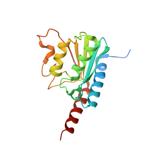
Crystal Structure of tRNA Adenosine Deaminase (TadA) from Aquifex aeolicus

Kuratani, M.Ishii, R.Bessho, Y.Fukunaga, R.Sengoku, T.Shirouzu, M.Sekine, S.Yokoyama, S.

(2005) J Biological Chem 280: 16002-16008

  • DOI: https://doi.org/10.1074/jbc.M414541200
  • Primary Citation Related Structures: 
    1WWR

  • PubMed Abstract: 

    The bacterial tRNA adenosine deaminase (TadA) generates inosine by deaminating the adenosine residue at the wobble position of tRNA(Arg-2). This modification is essential for the decoding system. In this study, we determined the crystal structure of Aquifex aeolicus TadA at a 1.8-A resolution. This is the first structure of a deaminase acting on tRNA. A. aeolicus TadA has an alpha/beta/alpha three-layered fold and forms a homodimer. The A. aeolicus TadA dimeric structure is completely different from the tetrameric structure of yeast CDD1, which deaminates mRNA and cytidine, but is similar to the dimeric structure of yeast cytosine deaminase. However, in the A. aeolicus TadA structure, the shapes of the C-terminal helix and the regions between the beta4 and beta5 strands are quite distinct from those of yeast cytosine deaminase and a large cavity is produced. This cavity contains many conserved amino acid residues that are likely to be involved in either catalysis or tRNA binding. We made a docking model of TadA with the tRNA anticodon stem loop.


  • Organizational Affiliation
    • Department of Biophysics and Biochemistry, Graduate School of Science, The University of Tokyo, 7-3-1 Hongo, Bunkyo-ku, Tokyo 113-0033, Japan.

Macromolecule Content 

  • Total Structure Weight: 78.04 kDa 
  • Atom Count: 5,495 
  • Modeled Residue Count: 622 
  • Deposited Residue Count: 684 
  • Unique protein chains: 1

Macromolecules

Find similar proteins by:|  3D Structure
Entity ID: 1
MoleculeChains  Sequence LengthOrganismDetailsImage
tRNA adenosine deaminase TadA
A, B, C, D
171Aquifex aeolicusMutation(s): 0 
EC: 3.5.4 (PDB Primary Data), 3.5.4.33 (UniProt)
UniProt
Find proteins for O67050 (Aquifex aeolicus (strain VF5))
Explore O67050 
Go to UniProtKB:  O67050
Entity Groups
Sequence Clusters30% Identity50% Identity70% Identity90% Identity95% Identity100% Identity
UniProt GroupO67050
Sequence Annotations
Expand
Reference Sequence

Experimental Data & Validation

Experimental Data

  • Method: X-RAY DIFFRACTION
  • Resolution: 1.80 Å
  • R-Value Free:  0.248 (Depositor), 0.239 (DCC) 
  • R-Value Work:  0.198 (Depositor), 0.199 (DCC) 
  • R-Value Observed: 0.198 (Depositor) 
Space Group: P 1 21 1
Unit Cell:
Length ( Å )Angle ( ˚ )
a = 43.176α = 90
b = 151.953β = 113.42
c = 54.133γ = 90
Software Package:
Software NamePurpose
CNSrefinement
HKL-2000data reduction
SCALEPACKdata scaling
MOLREPphasing

Structure Validation

View Full Validation Report


Entry History 

Revision History  (Full details and data files)

  • Version 1.0: 2005-02-01
    Type: Initial release
  • Version 1.1: 2008-04-30
    Changes: Version format compliance
  • Version 1.2: 2011-07-13
    Changes: Version format compliance
  • Version 1.3: 2023-10-25
    Changes: Data collection, Database references, Derived calculations, Refinement description