1WVN

Crystal Structure of domain 3 of human alpha polyC binding protein


Experimental Data Snapshot

  • Method: X-RAY DIFFRACTION
  • Resolution: 2.10 Å
  • R-Value Free: 0.248 
  • R-Value Work: 0.190 
  • R-Value Observed: 0.214 

Starting Model: experimental
View more details

wwPDB Validation   3D Report Full Report


This is version 1.5 of the entry. See complete history


Literature

Structure and RNA binding of the third KH domain of poly(C)-binding protein 1.

Sidiqi, M.Wilce, J.A.Vivian, J.P.Porter, C.J.Barker, A.Leedman, P.J.Wilce, M.C.J.

(2005) Nucleic Acids Res 33: 1213-1221

  • DOI: https://doi.org/10.1093/nar/gki265
  • Primary Citation of Related Structures:  
    1WVN

  • PubMed Abstract: 

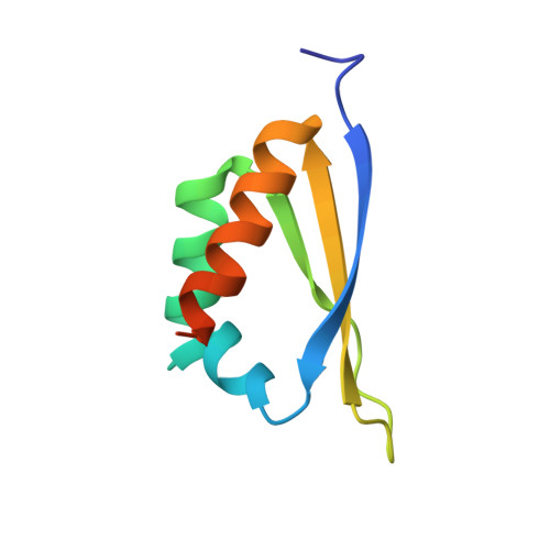
    Poly(C)-binding proteins (CPs) are important regulators of mRNA stability and translational regulation. They recognize C-rich RNA through their triple KH (hn RNP K homology) domain structures and are thought to carry out their function though direct protection of mRNA sites as well as through interactions with other RNA-binding proteins. We report the crystallographically derived structure of the third domain of alphaCP1 to 2.1 A resolution. alphaCP1-KH3 assumes a classical type I KH domain fold with a triple-stranded beta-sheet held against a three-helix cluster in a betaalphaalphabetabetaalpha configuration. Its binding affinity to an RNA sequence from the 3'-untranslated region (3'-UTR) of androgen receptor mRNA was determined using surface plasmon resonance, giving a K(d) of 4.37 microM, which is indicative of intermediate binding. A model of alphaCP1-KH3 with poly(C)-RNA was generated by homology to a recently reported RNA-bound KH domain structure and suggests the molecular basis for oligonucleotide binding and poly(C)-RNA specificity.


  • Organizational Affiliation

    School of Biomedical and Chemical Sciences, the UWA Centre for Medical Research, The University of Western Australia WA Australia 6009.


Macromolecules
Find similar proteins by:  (by identity cutoff)  |  3D Structure
Entity ID: 1
MoleculeChains Sequence LengthOrganismDetailsImage
Poly(rC)-binding protein 182Homo sapiensMutation(s): 0 
UniProt & NIH Common Fund Data Resources
Find proteins for Q15365 (Homo sapiens)
Explore Q15365 
Go to UniProtKB:  Q15365
GTEx:  ENSG00000169564 
Entity Groups  
Sequence Clusters30% Identity50% Identity70% Identity90% Identity95% Identity100% Identity
UniProt GroupQ15365
Sequence Annotations
Expand
  • Reference Sequence
Experimental Data & Validation

Experimental Data

  • Method: X-RAY DIFFRACTION
  • Resolution: 2.10 Å
  • R-Value Free: 0.248 
  • R-Value Work: 0.190 
  • R-Value Observed: 0.214 
  • Space Group: P 21 21 2
Unit Cell:
Length ( Å )Angle ( ˚ )
a = 33.4α = 90
b = 71β = 90
c = 29.1γ = 90
Software Package:
Software NamePurpose
REFMACrefinement
SCALEPACKdata scaling
AMoREphasing

Structure Validation

View Full Validation Report


Entry History 

Deposition Data

Revision History  (Full details and data files)

  • Version 1.0: 2005-04-05
    Type: Initial release
  • Version 1.1: 2008-04-30
    Changes: Version format compliance
  • Version 1.2: 2011-07-13
    Changes: Version format compliance
  • Version 1.3: 2017-10-11
    Changes: Refinement description
  • Version 1.4: 2019-01-23
    Changes: Data collection, Structure summary
  • Version 1.5: 2023-10-25
    Changes: Data collection, Database references, Refinement description