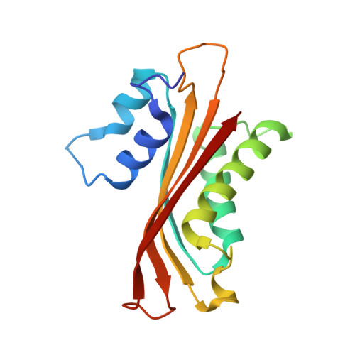
1WRN

Metal Ion dependency of the antiterminator protein, HutP, for binding to the terminator region of hut mRNA- A structural basis


Experimental Data Snapshot

  • Method: X-RAY DIFFRACTION
  • Resolution: 2.30 Å
  • R-Value Free: 0.305 
  • R-Value Work: 0.254 
  • R-Value Observed: 0.254 

wwPDB Validation   3D Report Full Report


Ligand Structure Quality Assessment 


This is version 1.4 of the entry. See complete history


Literature

Characterization of the metal ion binding site in the anti-terminator protein, HutP, of Bacillus subtilis

Kumarevel, T.Mizuno, H.Kumar, P.K.R.

(2005) Nucleic Acids Res 33: 5494-5502

  • DOI: https://doi.org/10.1093/nar/gki868
  • Primary Citation of Related Structures:  
    1WPT, 1WRN, 1WRO

  • PubMed Abstract: 

    HutP is an RNA-binding protein that regulates the expression of the histidine utilization (hut) operon in Bacillus subtilis, by binding to cis-acting regulatory sequences on hut mRNA. It requires L-histidine and an Mg2+ ion for binding to the specific sequence within the hut mRNA. In the present study, we show that several divalent cations can mediate the HutP-RNA interactions. The best divalent cations were Mn2+, Zn2+ and Cd2+, followed by Mg2+, Co2+ and Ni2+, while Cu2+, Yb2+ and Hg2+ were ineffective. In the HutP-RNA interactions, divalent cations cannot be replaced by monovalent cations, suggesting that a divalent metal ion is required for mediating the protein-RNA interactions. To clarify their importance, we have crystallized HutP in the presence of three different metal ions (Mg2+, Mn2+ and Ba2+), which revealed the importance of the metal ion binding site. Furthermore, these analyses clearly demonstrated how the metal ions cause the structural rearrangements that are required for the hut mRNA recognition.


  • Organizational Affiliation

    Institute for Biological Resources and Functions, National Institute of Advanced Industrial Science and Technology (AIST) Central 6, Tsukuba, Ibaraki 305-8566, Japan.


Macromolecules
Find similar proteins by:  (by identity cutoff)  |  3D Structure
Entity ID: 1
MoleculeChains Sequence LengthOrganismDetailsImage
Hut operon positive regulatory protein
A, B, C
147Bacillus subtilisMutation(s): 1 
UniProt
Find proteins for P10943 (Bacillus subtilis (strain 168))
Explore P10943 
Go to UniProtKB:  P10943
Entity Groups  
Sequence Clusters30% Identity50% Identity70% Identity90% Identity95% Identity100% Identity
UniProt GroupP10943
Sequence Annotations
Expand
  • Reference Sequence
Small Molecules
Ligands 3 Unique
IDChains Name / Formula / InChI Key2D Diagram3D Interactions
HIS
Query on HIS

Download Ideal Coordinates CCD File 
E [auth A],
K [auth B],
N [auth C]
HISTIDINE
C6 H10 N3 O2
HNDVDQJCIGZPNO-YFKPBYRVSA-O
PEG
Query on PEG

Download Ideal Coordinates CCD File 
F [auth A]
G [auth A]
H [auth A]
I [auth A]
L [auth B]
F [auth A],
G [auth A],
H [auth A],
I [auth A],
L [auth B],
O [auth C]
DI(HYDROXYETHYL)ETHER
C4 H10 O3
MTHSVFCYNBDYFN-UHFFFAOYSA-N
MN
Query on MN

Download Ideal Coordinates CCD File 
D [auth A],
J [auth B],
M [auth C]
MANGANESE (II) ION
Mn
WAEMQWOKJMHJLA-UHFFFAOYSA-N
Experimental Data & Validation

Experimental Data

  • Method: X-RAY DIFFRACTION
  • Resolution: 2.30 Å
  • R-Value Free: 0.305 
  • R-Value Work: 0.254 
  • R-Value Observed: 0.254 
  • Space Group: P 21 21 2
Unit Cell:
Length ( Å )Angle ( ˚ )
a = 77.76α = 90
b = 81.4β = 90
c = 76.04γ = 90
Software Package:
Software NamePurpose
CNSrefinement
HKL-2000data reduction
CCP4data scaling
CNSphasing

Structure Validation

View Full Validation Report


Ligand Structure Quality Assessment 


Entry History 

Deposition Data

Revision History  (Full details and data files)

  • Version 1.0: 2005-08-30
    Type: Initial release
  • Version 1.1: 2008-04-30
    Changes: Version format compliance
  • Version 1.2: 2011-07-13
    Changes: Derived calculations, Version format compliance
  • Version 1.3: 2021-11-10
    Changes: Database references, Derived calculations
  • Version 1.4: 2024-05-29
    Changes: Data collection