1WRL | pdb_00001wrl

Crystal structure of the N-terminal domain of human cardiac troponin C in complex with trifluoperazine (monoclinic crystal form)


Experimental Data Snapshot

  • Method: X-RAY DIFFRACTION
  • Resolution: 2.60 Å
  • R-Value Free: 
    0.273 (Depositor), 0.267 (DCC) 
  • R-Value Work: 
    0.240 (Depositor), 0.234 (DCC) 
  • R-Value Observed: 
    0.242 (Depositor) 

wwPDB Validation 3D Report Full Report

Validation slider image for 1WRL

Ligand Structure Quality Assessment 


This is version 1.4 of the entry. See complete history

Literature

Crystal structure of the N-terminal domain of human cardiac troponin C in complex with trifluoperazine

Takeda, S.Igarashi, T.Oishi, Y.Mori, H.

To be published.

Macromolecule Content 

  • Total Structure Weight: 64.84 kDa 
  • Atom Count: 4,301 
  • Modeled Residue Count: 495 
  • Deposited Residue Count: 528 
  • Unique protein chains: 1

Macromolecules

Find similar proteins by:|  3D Structure
Entity ID: 1
MoleculeChains  Sequence LengthOrganismDetailsImage
Troponin C, slow skeletal and cardiac muscles
A, B, C, D, E
A, B, C, D, E, F
88Homo sapiensMutation(s): 2 
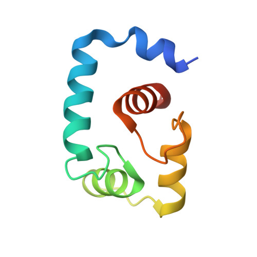
UniProt & NIH Common Fund Data Resources
Find proteins for P63316 (Homo sapiens)
Explore P63316 
Go to UniProtKB:  P63316
PHAROS:  P63316
GTEx:  ENSG00000114854 
Entity Groups
Sequence Clusters30% Identity50% Identity70% Identity90% Identity95% Identity100% Identity
UniProt GroupP63316
Sequence Annotations
Expand
Reference Sequence

Small Molecules

Ligands 2 Unique
IDChains Name / Formula / InChI Key2D Diagram3D Interactions
TFP

Query on TFP


Download:Ideal Coordinates CCD File
H [auth A]
I [auth A]
K [auth B]
L [auth B]
N [auth C]
H [auth A],
I [auth A],
K [auth B],
L [auth B],
N [auth C],
O [auth C],
Q [auth D],
R [auth D],
T [auth E],
U [auth E],
W [auth F],
X [auth F]
10-[3-(4-METHYL-PIPERAZIN-1-YL)-PROPYL]-2-TRIFLUOROMETHYL-10H-PHENOTHIAZINE
C21 H24 F3 N3 S
ZEWQUBUPAILYHI-UHFFFAOYSA-N
CA

Query on CA


Download:Ideal Coordinates CCD File
G [auth A]
J [auth B]
M [auth C]
P [auth D]
S [auth E]
G [auth A],
J [auth B],
M [auth C],
P [auth D],
S [auth E],
V [auth F]
CALCIUM ION
Ca
BHPQYMZQTOCNFJ-UHFFFAOYSA-N

Experimental Data & Validation

Experimental Data

  • Method: X-RAY DIFFRACTION
  • Resolution: 2.60 Å
  • R-Value Free:  0.273 (Depositor), 0.267 (DCC) 
  • R-Value Work:  0.240 (Depositor), 0.234 (DCC) 
  • R-Value Observed: 0.242 (Depositor) 
Space Group: P 1 21 1
Unit Cell:
Length ( Å )Angle ( ˚ )
a = 48.93α = 90
b = 67.46β = 96.75
c = 90.28γ = 90
Software Package:
Software NamePurpose
HKL-2000data collection
SCALEPACKdata scaling
MOLREPphasing
CNSrefinement
HKL-2000data reduction

Structure Validation

View Full Validation Report


Ligand Structure Quality Assessment 


Entry History 

Deposition Data

Revision History  (Full details and data files)

  • Version 1.0: 2006-01-24
    Type: Initial release
  • Version 1.1: 2008-04-30
    Changes: Version format compliance
  • Version 1.2: 2011-07-13
    Changes: Version format compliance
  • Version 1.3: 2021-11-10
    Changes: Database references, Derived calculations
  • Version 1.4: 2024-05-29
    Changes: Data collection