1WQ5 | pdb_00001wq5

Crystal structure of tryptophan synthase alpha-subunit from Escherichia coli


Experimental Data Snapshot

  • Method: X-RAY DIFFRACTION
  • Resolution: 2.30 Å
  • R-Value Free: 
    0.248 (Depositor), 0.179 (DCC) 
  • R-Value Work: 
    0.185 (Depositor), 0.190 (DCC) 
  • R-Value Observed: 
    0.185 (Depositor) 

Starting Model: experimental
View more details

wwPDB Validation 3D Report Full Report

Validation slider image for 1WQ5

This is version 1.4 of the entry. See complete history

Literature

Conformational Changes in the alpha-Subunit Coupled to Binding of the beta(2)-Subunit of Tryptophan Synthase from Escherichia coli: Crystal Structure of the Tryptophan Synthase alpha-Subunit Alon

Nishio, K.Morimoto, Y.Ishizuka, M.Ogasahara, K.Tsukihara, T.Yutani, K.

(2005) Biochemistry 44: 1184-1192

  • DOI: https://doi.org/10.1021/bi047927m
  • Primary Citation Related Structures: 
    1V7Y, 1WQ5

  • PubMed Abstract: 

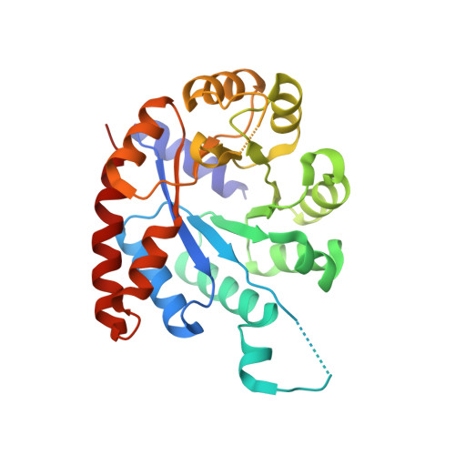
    When the tryptophan synthase alpha- and beta(2)-subunits combine to form the alpha(2)beta(2)-complex, the enzymatic activity of each subunit is stimulated by 1-2 orders of magnitude. To elucidate the structural basis of this mutual activation, it is necessary to determine the structures of the alpha- and beta-subunits alone and together with the alpha(2)beta(2)-complex. The crystal structures of the tryptophan synthase alpha(2)beta(2)-complex from Salmonella typhimurium (Stalpha(2)beta(2)-complex) have already been reported. However, the structures of the subunit alone from mesophiles have not yet been determined. The structure of the tryptophan synthase alpha-subunit alone from Escherichia coli (Ecalpha-subunit) was determined by an X-ray crystallographic analysis at 2.3 A, which is the first report on the subunits alone from the mesophiles. The biggest difference between the structures of the Ecalpha-subunit alone and the alpha-subunit in the Stalpha(2)beta(2)-complex (Stalpha-subunit) was as follows. Helix 2' in the Stalpha-subunit, including an active site residue (Asp60), was changed to a flexible loop in the Ecalpha-subunit alone. The conversion of the helix to a loop resulted in the collapse of the correct active site conformation. This region is also an important part for the mutual activation in the Stalpha(2)beta(2)-complex and interaction with the beta-subunit. These results suggest that the formation of helix 2'that is essential for the stimulation of the enzymatic activity of the alpha-subunit is constructed by the induced-fit mode involved in conformational changes upon interaction between the alpha- and beta-subunits. This also confirms the prediction of the conformational changes based on the thermodynamic analysis for the association between the alpha- and beta-subunits.


  • Organizational Affiliation
    • Institute for Protein Research, Osaka University, 3-2 Yamadaoka, Suita, Osaka 565-0871, Japan.

Macromolecule Content 

  • Total Structure Weight: 59.49 kDa 
  • Atom Count: 4,264 
  • Modeled Residue Count: 519 
  • Deposited Residue Count: 536 
  • Unique protein chains: 1

Macromolecules

Find similar proteins by:|  3D Structure
Entity ID: 1
MoleculeChains  Sequence LengthOrganismDetailsImage
Tryptophan synthase alpha chain
A, B
268Escherichia coliMutation(s): 0 
EC: 4.2.1.20
UniProt
Find proteins for P0A877 (Escherichia coli (strain K12))
Explore P0A877 
Go to UniProtKB:  P0A877
Entity Groups
Sequence Clusters30% Identity50% Identity70% Identity90% Identity95% Identity100% Identity
UniProt GroupP0A877
Sequence Annotations
Expand
Reference Sequence

Small Molecules

Ligands 2 Unique
IDChains Name / Formula / InChI Key2D Diagram3D Interactions
SO4

Query on SO4


Download:Ideal Coordinates CCD File
C [auth A]
D [auth A]
E [auth A]
F [auth A]
G [auth A]
C [auth A],
D [auth A],
E [auth A],
F [auth A],
G [auth A],
H [auth A],
I [auth A],
P [auth B],
Q [auth B],
R [auth B],
S [auth B],
T [auth B],
U [auth B]
SULFATE ION
O4 S
QAOWNCQODCNURD-UHFFFAOYSA-L
GOL

Query on GOL


Download:Ideal Coordinates CCD File
J [auth A]
K [auth A]
L [auth A]
M [auth A]
N [auth A]
J [auth A],
K [auth A],
L [auth A],
M [auth A],
N [auth A],
O [auth A],
V [auth B],
W [auth B]
GLYCEROL
C3 H8 O3
PEDCQBHIVMGVHV-UHFFFAOYSA-N

Experimental Data & Validation

Experimental Data

  • Method: X-RAY DIFFRACTION
  • Resolution: 2.30 Å
  • R-Value Free:  0.248 (Depositor), 0.179 (DCC) 
  • R-Value Work:  0.185 (Depositor), 0.190 (DCC) 
  • R-Value Observed: 0.185 (Depositor) 
Space Group: C 1 2 1
Unit Cell:
Length ( Å )Angle ( ˚ )
a = 156.774α = 90
b = 44.626β = 96.46
c = 71.716γ = 90
Software Package:
Software NamePurpose
CNSrefinement
DENZOdata reduction
SCALEPACKdata scaling
CNSphasing

Structure Validation

View Full Validation Report


Entry History 

Revision History  (Full details and data files)

  • Version 1.0: 2005-02-15
    Type: Initial release
  • Version 1.1: 2008-04-30
    Changes: Version format compliance
  • Version 1.2: 2011-07-13
    Changes: Derived calculations, Version format compliance
  • Version 1.3: 2024-03-13
    Changes: Data collection, Database references, Derived calculations
  • Version 1.4: 2024-04-03
    Changes: Refinement description