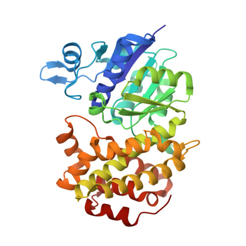
1WPQ | pdb_00001wpq

Ternary Complex Of Glycerol 3-phosphate Dehydrogenase 1 with NAD and dihydroxyactone


Experimental Data Snapshot

  • Method: X-RAY DIFFRACTION
  • Resolution: 2.50 Å
  • R-Value Free: 
    0.294 (Depositor), 0.280 (DCC) 
  • R-Value Work: 
    0.258 (Depositor), 0.250 (DCC) 
  • R-Value Observed: 
    0.263 (Depositor) 

wwPDB Validation   3D Report Full Report


Ligand Structure Quality Assessment 

Created with Raphaël 2.3.0Worse 01 BetterLigand structure goodness of fit to experimental dataBest fitted NADClick on this verticalbar to view detailsBest fitted 13PClick on this verticalbar to view details

This is version 1.3 of the entry. See complete history


Literature

Crystal Structures of Human Glycerol 3-phosphate Dehydrogenase 1 (GPD1)

Ou, X.Ji, C.Han, X.Zhao, X.Li, X.Mao, Y.Wong, L.L.Bartlam, M.Rao, Z.

(2006) J Mol Biology 357: 858-869

  • DOI: https://doi.org/10.1016/j.jmb.2005.12.074
  • Primary Citation of Related Structures:  
    1WPQ, 1X0V, 1X0X

  • PubMed Abstract: 

    Homo sapiens L-alpha-glycerol-3-phosphate dehydrogenase 1 (GPD1) catalyzes the reversible biological conversion of dihydroxyacetone (DHAP) to glycerol-3-phosphate. The GPD1 protein was expressed in Escherichia coli, and purified as a fusion protein with glutathione S-transferase. Here we report the apoenzyme structure of GPD1 determined by multiwavelength anomalous diffraction phasing, and other complex structures with small molecules (NAD+ and DHAP) by the molecular replacement method. This enzyme structure is organized into two distinct domains, the N-terminal eight-stranded beta-sheet sandwich domain and the C-terminal helical substrate-binding domain. An electrophilic catalytic mechanism by the epsilon-NH3+ group of Lys204 is proposed on the basis of the structural analyses. In addition, the inhibitory effects of zinc and sulfate on GPDHs are assayed and discussed.


  • Organizational Affiliation

    National Laboratory of Biomacromolecules, Institute of Biophysics (IBP), Chinese Academy of Sciences, Beijing 100101, China.


Macromolecules
Find similar proteins by:  (by identity cutoff)  |  3D Structure
Entity ID: 1
MoleculeChains Sequence LengthOrganismDetailsImage
Glycerol-3-phosphate dehydrogenase [NAD+], cytoplasmic
A, B
349Homo sapiensMutation(s): 0 
EC: 1.1.1.8
UniProt & NIH Common Fund Data Resources
Find proteins for P21695 (Homo sapiens)
Explore P21695 
Go to UniProtKB:  P21695
PHAROS:  P21695
GTEx:  ENSG00000167588 
Entity Groups  
Sequence Clusters30% Identity50% Identity70% Identity90% Identity95% Identity100% Identity
UniProt GroupP21695
Sequence Annotations
Expand
  • Reference Sequence
Small Molecules
Ligands 3 Unique
IDChains Name / Formula / InChI Key2D Diagram3D Interactions
NAD
Query on NAD

Download Ideal Coordinates CCD File 
J [auth A],
M [auth B]
NICOTINAMIDE-ADENINE-DINUCLEOTIDE
C21 H27 N7 O14 P2
BAWFJGJZGIEFAR-NNYOXOHSSA-N
13P
Query on 13P

Download Ideal Coordinates CCD File 
I [auth A],
L [auth B]
1,3-DIHYDROXYACETONEPHOSPHATE
C3 H7 O6 P
GNGACRATGGDKBX-UHFFFAOYSA-N
SO4
Query on SO4

Download Ideal Coordinates CCD File 
C [auth A]
D [auth A]
E [auth A]
F [auth A]
G [auth A]
SULFATE ION
O4 S
QAOWNCQODCNURD-UHFFFAOYSA-L
Experimental Data & Validation

Experimental Data

  • Method: X-RAY DIFFRACTION
  • Resolution: 2.50 Å
  • R-Value Free:  0.294 (Depositor), 0.280 (DCC) 
  • R-Value Work:  0.258 (Depositor), 0.250 (DCC) 
  • R-Value Observed: 0.263 (Depositor) 
Space Group: P 43 21 2
Unit Cell:
Length ( Å )Angle ( ˚ )
a = 115.995α = 90
b = 115.995β = 90
c = 153.72γ = 90
Software Package:
Software NamePurpose
DENZOdata reduction
SCALEPACKdata scaling
CNSrefinement
CNSphasing

Structure Validation

View Full Validation Report


Ligand Structure Quality Assessment 

Created with Raphaël 2.3.0Worse 01 BetterLigand structure goodness of fit to experimental dataBest fitted NADClick on this verticalbar to view detailsBest fitted 13PClick on this verticalbar to view details

Entry History 

Deposition Data

Revision History  (Full details and data files)

  • Version 1.0: 2006-04-11
    Type: Initial release
  • Version 1.1: 2008-04-30
    Changes: Version format compliance
  • Version 1.2: 2011-07-13
    Changes: Version format compliance
  • Version 1.3: 2024-03-13
    Changes: Data collection, Database references, Derived calculations