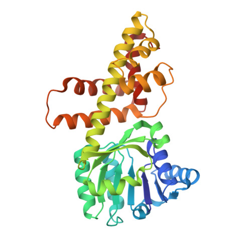
1WP4

Structure of TT368 protein from Thermus Thermophilus HB8


Experimental Data Snapshot

  • Method: X-RAY DIFFRACTION
  • Resolution: 2.00 Å
  • R-Value Free: 0.208 
  • R-Value Work: 0.182 
  • R-Value Observed: 0.182 

Starting Model: experimental
View more details

wwPDB Validation   3D Report Full Report


Ligand Structure Quality Assessment 


This is version 1.3 of the entry. See complete history


Literature

Crystal Structure of Novel NADP-dependent 3-Hydroxyisobutyrate Dehydrogenase from Thermus thermophilus HB8

Lokanath, N.K.Ohshima, N.Takio, K.Shiromizu, I.Kuroishi, C.Okazaki, N.Kuramitsu, S.Yokoyama, S.Miyano, M.Kunishima, N.

(2005) J Mol Biol 352: 905-917

  • DOI: https://doi.org/10.1016/j.jmb.2005.07.068
  • Primary Citation of Related Structures:  
    1WP4, 2CVZ

  • PubMed Abstract: 

    3-Hydroxyisobutyrate, a central metabolite in the valine catabolic pathway, is reversibly oxidized to methylmalonate semialdehyde by a specific dehydrogenase belonging to the 3-hydroxyacid dehydrogenase family. To gain insight into the function of this enzyme at the atomic level, we have determined the first crystal structures of the 3-hydroxyisobutyrate dehydrogenase from Thermus thermophilus HB8: holo enzyme and sulfate ion complex. The crystal structures reveal a unique tetrameric oligomerization and a bound cofactor NADP+. This bacterial enzyme may adopt a novel cofactor-dependence on NADP, whereas NAD is preferred in eukaryotic enzymes. The protomer folds into two distinct domains with open/closed interdomain conformations. The cofactor NADP+ with syn nicotinamide and the sulfate ion are bound to distinct sites located at the interdomain cleft of the protomer through an induced-fit domain closure upon cofactor binding. From the structural comparison with the crystal structure of 6-phosphogluconate dehydrogenase, another member of the 3-hydroxyacid dehydrogenase family, it is suggested that the observed sulfate ion and the substrate 3-hydroxyisobutyrate share the same binding pocket. The observed oligomeric state might be important for the catalytic function through forming the active site involving two adjacent subunits, which seems to be conserved in the 3-hydroxyacid dehydrogenases. A kinetic study confirms that this enzyme has strict substrate specificity for 3-hydroxyisobutyrate and serine, but it cannot distinguish the chirality of the substrates. Lys165 is likely the catalytic residue of the enzyme.


  • Organizational Affiliation

    Highthroughput Factory, RIKEN Harima Institute at SPring-8, 1-1-1 Kouto, Mikazuki-cho, Sayo-gun, Hyogo 679-5148, Japan.


Macromolecules
Find similar proteins by:  (by identity cutoff)  |  3D Structure
Entity ID: 1
MoleculeChains Sequence LengthOrganismDetailsImage
3-hydroxyisobutyrate dehydrogenase
A, B, C, D
289Thermus thermophilus HB8Mutation(s): 0 
EC: 1.1.1.31
UniProt
Find proteins for Q5SLQ6 (Thermus thermophilus (strain ATCC 27634 / DSM 579 / HB8))
Explore Q5SLQ6 
Go to UniProtKB:  Q5SLQ6
Entity Groups  
Sequence Clusters30% Identity50% Identity70% Identity90% Identity95% Identity100% Identity
UniProt GroupQ5SLQ6
Sequence Annotations
Expand
  • Reference Sequence
Small Molecules
Ligands 2 Unique
IDChains Name / Formula / InChI Key2D Diagram3D Interactions
NDP
Query on NDP

Download Ideal Coordinates CCD File 
F [auth A],
H [auth B],
J [auth C],
L [auth D]
NADPH DIHYDRO-NICOTINAMIDE-ADENINE-DINUCLEOTIDE PHOSPHATE
C21 H30 N7 O17 P3
ACFIXJIJDZMPPO-NNYOXOHSSA-N
SO4
Query on SO4

Download Ideal Coordinates CCD File 
E [auth A],
G [auth B],
I [auth C],
K [auth D]
SULFATE ION
O4 S
QAOWNCQODCNURD-UHFFFAOYSA-L
Experimental Data & Validation

Experimental Data

  • Method: X-RAY DIFFRACTION
  • Resolution: 2.00 Å
  • R-Value Free: 0.208 
  • R-Value Work: 0.182 
  • R-Value Observed: 0.182 
  • Space Group: P 21 21 21
Unit Cell:
Length ( Å )Angle ( ˚ )
a = 93.863α = 90
b = 109.028β = 90
c = 116.392γ = 90
Software Package:
Software NamePurpose
CNSrefinement
HKL-2000data reduction
SCALEPACKdata scaling
EPMRphasing

Structure Validation

View Full Validation Report


Ligand Structure Quality Assessment 


Entry History 

Deposition Data

Revision History  (Full details and data files)

  • Version 1.0: 2005-08-30
    Type: Initial release
  • Version 1.1: 2008-04-30
    Changes: Version format compliance
  • Version 1.2: 2011-07-13
    Changes: Source and taxonomy, Version format compliance
  • Version 1.3: 2023-10-25
    Changes: Data collection, Database references, Derived calculations, Refinement description