1WLE | pdb_00001wle

Crystal Structure of mammalian mitochondrial seryl-tRNA synthetase complexed with seryl-adenylate


Experimental Data Snapshot

  • Method: X-RAY DIFFRACTION
  • Resolution: 1.65 Å
  • R-Value Free: 
    0.226 (Depositor), 0.206 (DCC) 
  • R-Value Work: 
    0.210 (Depositor), 0.212 (DCC) 

Starting Model: experimental
View more details

wwPDB Validation 3D Report Full Report

Validation slider image for 1WLE

Ligand Structure Quality Assessment 


This is version 1.3 of the entry. See complete history

Literature

Dual-mode recognition of noncanonical tRNAs(Ser) by seryl-tRNA synthetase in mammalian mitochondria

Chimnaronk, S.Jeppesen, M.G.Suzuki, T.Nyborg, J.Watanabe, K.

(2005) EMBO J 24: 3369-3379

  • DOI: https://doi.org/10.1038/sj.emboj.7600811
  • Primary Citation Related Structures: 
    1WLE

  • PubMed Abstract: 

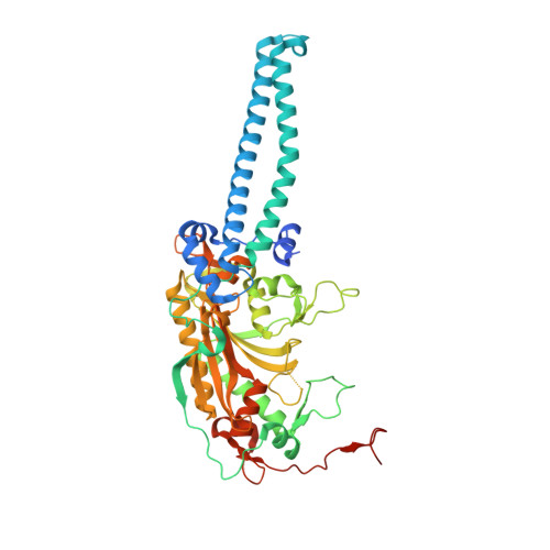
    The secondary structures of metazoan mitochondrial (mt) tRNAs(Ser) deviate markedly from the paradigm of the canonical cloverleaf structure; particularly, tRNA(Ser)(GCU) corresponding to the AGY codon (Y=U and C) is highly truncated and intrinsically missing the entire dihydrouridine arm. None of the mt serine isoacceptors possesses the elongated variable arm, which is the universal landmark for recognition by seryl-tRNA synthetase (SerRS). Here, we report the crystal structure of mammalian mt SerRS from Bos taurus in complex with seryl adenylate at an atomic resolution of 1.65 A. Coupling structural information with a tRNA-docking model and the mutagenesis studies, we have unraveled the key elements that establish tRNA binding specificity, differ from all other known bacterial and eukaryotic systems, are the characteristic extensions in both extremities, as well as a few basic residues residing in the amino-terminal helical arm of mt SerRS. Our data further uncover an unprecedented mechanism of a dual-mode recognition employed to discriminate two distinct 'bizarre' mt tRNAs(Ser) by alternative combination of interaction sites.


  • Organizational Affiliation
    • Department of Integrated Biosciences, Graduate School of Frontier Sciences, University of Tokyo, Kashiwanoha, Kashiwa, Chiba, Japan.

Macromolecule Content 

  • Total Structure Weight: 113.99 kDa 
  • Atom Count: 8,106 
  • Modeled Residue Count: 934 
  • Deposited Residue Count: 1,002 
  • Unique protein chains: 1

Macromolecules

Find similar proteins by:|  3D Structure
Entity ID: 1
MoleculeChains  Sequence LengthOrganismDetailsImage
Seryl-tRNA synthetase
A, B
501Bos taurusMutation(s): 0 
Gene Names: SerRSmt
EC: 6.1.1.11
UniProt
Find proteins for Q9N0F3 (Bos taurus)
Explore Q9N0F3 
Go to UniProtKB:  Q9N0F3
Entity Groups
Sequence Clusters30% Identity50% Identity70% Identity90% Identity95% Identity100% Identity
UniProt GroupQ9N0F3
Sequence Annotations
Expand
Reference Sequence

Experimental Data & Validation

Experimental Data

  • Method: X-RAY DIFFRACTION
  • Resolution: 1.65 Å
  • R-Value Free:  0.226 (Depositor), 0.206 (DCC) 
  • R-Value Work:  0.210 (Depositor), 0.212 (DCC) 
Space Group: C 2 2 21
Unit Cell:
Length ( Å )Angle ( ˚ )
a = 79.864α = 90
b = 230.35β = 90
c = 135.59γ = 90
Software Package:
Software NamePurpose
DENZOdata reduction
SCALEPACKdata scaling
AMoREphasing
CNSrefinement

Structure Validation

View Full Validation Report


Ligand Structure Quality Assessment 


Entry History 

Deposition Data

Revision History  (Full details and data files)

  • Version 1.0: 2005-09-06
    Type: Initial release
  • Version 1.1: 2008-04-30
    Changes: Version format compliance
  • Version 1.2: 2011-07-13
    Changes: Version format compliance
  • Version 1.3: 2023-10-25
    Changes: Data collection, Database references, Derived calculations, Refinement description